
- Similar to other vertebrates, the nervous system of frog is composed of 3 main sub-divisions:
I. Central Nervous system of frog
1. Brain of Frog:
- Brain is white in color, elongated and somewhat flattened structure.
- It lies well protected inside the cranium of skull.
- It is surrounded by two meninges.
- Meninges are connective tissue membranes.
- The inner or piamater is delicate and pigmented.
- Piamater is vascular and closely applied to brain.
- The outer durameter is tough and fibrous which lines the cranial cavity.
- The narrow space between the membranes and the inner cavities of brain are filled with cerebrospinal fluid.
- Cerebrospinal fluid is a clear, watery and lymphatic fluid. It protects and nourishes the brain.
- The ventricles (inner cavities of brain) are continuous with one another.
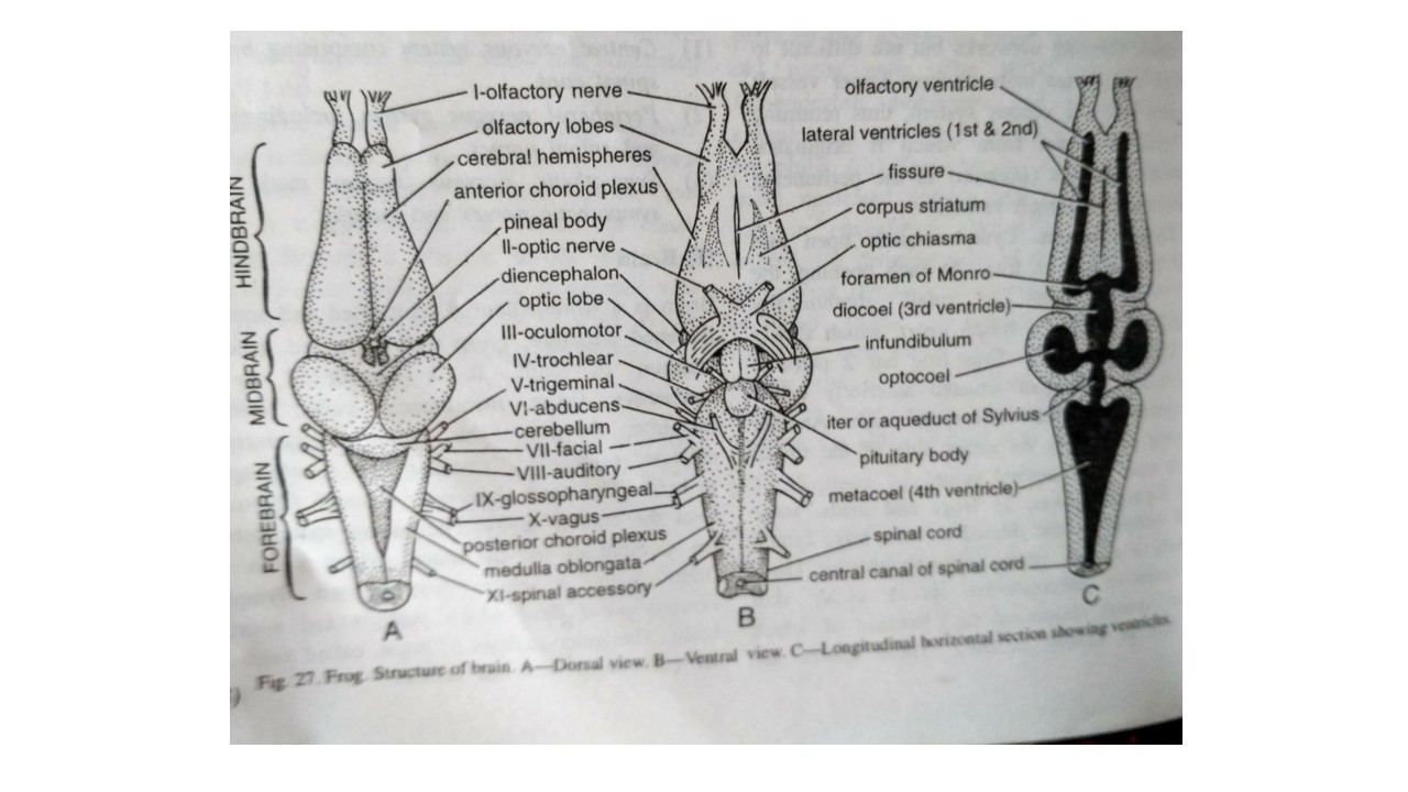
Brain can be explained under 3 main parts:
i. Forebrain:
- It consists of two olfactory lobes, two cerebral hemispheres and a diencephalon.
- Olfactory lobes:
- Olfactory lobes are two anterior most, rather small and spherical lobes.
- Each lobe sends a small olfactory nerve to the nasal chamber of its side.
- The two lobes are united but each contains a separate small cavity.
- This cavity is termed as the olfactory ventricle or rhinocoel.
- Olfactory lobes are concerned with sense of smell which is poorly developed in frog.
- Cerebral hemispheres:
- Posteriorly, the olfactory lobes are demarcated by a slight constriction from two large cerebral hemispheres that forms cerebrum.
- Cerebral hemispheres are long, oval, and smooth structures.
- It is narrow in front but broad behind.
- It is separated from one another by a deep mid-longitudinal groove or fissure.
- The large cavities of hemispheres, called I, II, or lateral ventricles or paracoels, are continuous anteriorly with olfactory ventricles.
- Posteriorly they unite with each other and with the III ventricle or diocoel of diencephalon through foramen of Monro.
- Foramen of Monro is the common opening.
- The roof of cerebrum is thin and is called as pallium.
- The each ventro-lateral side is thick called as corpus striatum (singular).
- Corpus striatum is made of white medullated nerve fibers and cells.
- A transverse fibrous tract, the anterior commissure joins the two corpora striata (pleural) together.
- The hemispheres are the place of memory, intelligence, consciousness and will.
- It also regulates voluntary actions.
- Diencephalon:
- It is the short, rhomboid, depressed region.
- It lies just behind the cerebrum.
- Its small cavity is termed as third ventricle or diocoel.
- Diocoel has thick lateral walls called as optic thalamic and a thick floor, called as hypothalamus.
- Its dorsal roof contains a network of blood capillaries, called as anterior choroid plexus.
- Behind anterior choroid plexus, arises a small hollow projection, the pineal stalk, that runs to the brow spot.
- In case of tadpoles, a small spherical pineal body is attached to the stalk.
- However, in adult frog, it is detached and comes to lie outside skull.
- The two optic nerves when crossed, forms the X-shaped optic chiasma.
- It lies on the ventral surface of diencephalon.
- A large median bilobed projection, the infundibulum is present just posterior to optic chiasma.
- The infundibulum bears a flattened oval pituitary body or hypophysis.
- Diencephalon is concerned with vision and balance.
ii. Midbrain:
- It is the broadest part of brain.
- It consists of a narrow canal, called as iter or aqueduct of Sylvius, which is continuous with III ventricle in front and IV ventricle behind.
- Two large rounded optic lobes, called copro bigemina are present dorsolaterally in midbrain.
- Their canals called as optocoels open into iter.
- There are two thick longitudinal bands of nerve fibers, called as crura cerebri.
- They run longitudinally beneath optic lobes connecting diencephalon and medulla.
- The inhibition of spinal reflexes is done by optic lobes, and each lobe controls the opposite side of body.
iii. Hindbrain:
- It is the posterior part of brain.
- It includes cerebellum and medulla oblongata.
- Cerebellum:
- It is a poorly developed narrow ridge or band.
- It is placed dorsally just behind the optic lobes.
- It controls equilibrium and muscular co-ordination which are not that important in case of frog.
- Medulla oblongata:
- It is the last small part of brain.
- It is continuous with spinal cord without distinction.
- It has triangular cavity which is called as IV ventricle or metacoel.
- Metacoel is continuous anteriorly with iter and posteriorly with the central cavity of spinal cord.
- Metacoel has thin and highly vascular dorsal roof which forms the posterior choroid plexus.
- Medulla regulates essential involuntary functions as such heartbeat, metabolism, respiration, etc. which keeps on going even if rest of the brain is removed.
- However, the removal of medulla is soon followed by death.
2. Spinal cord of frog:
- Posteriorly the spinal cord extends from medulla oblongata through foramen magnum.
- It lies protected within the neural canal of vertebral column.
- It is short, thick, cylindrical, somewhat flattened and white in color.
- Similar to brain, spinal cord is also surrounded by the two meninges, piamater and duramater that contains protective and nourishing cerebrospinal fluid.
- Posteriorly, it ends into a fine, non-nervous filament, called as filum terminale in the urostyle.
- Spinal cord shows swelling in two places:
- Brachial enlargement: between forelimbs.
- Sciatic or lumbar swelling: anterior to filum terminale.
- Two longitudinal grooves run throughout the length of spinal cord.
- The mid-dorsal called as dorsal fissure and the mid-ventral is termed as ventral fissure.
- It encloses a narrow central canal which is a continuation of the ventricles of brain.
- Spinal cord comprises of:
- Outer white matter: composed mainly of nerve fibres.
- Inner gray matter: composed largely of nerve cells.
- Spinal cord is chiefly concerned with the reflex actions.
II. Peripheral nervous system of frog
i. Cranial nerves:
- From the brain of frog, 10 pairs of cranial nerves are originated.
- Some claim to have O or terminal nerves making that count to 11 pairs.
- Their number, name, origin, distribution and nature are shortly enlisted in the following table:
| Number | Name of cranial nerve | Origin | Distribution | Nature |
| O | Terminal | Forebrain | Lining of nose | Sensory(probably) |
| I | Olfactory | Olfactory lobe | Lining of nose | Sensory(small) |
| II | Optic | Diencephalon | Retina of eye | Sensory(vision) |
| III | Oculomotor | Midbrain ventrally | 4 muscles of eye | Motor |
| IV | Trochlear | Midbrain dorsally | Superior oblique muscle of eye | Motor |
| V | Trigeminal Ophthalmic Maxillary Mandibular | Medulla laterally | Skin of snout Skin of upper jaw Muscles of lower jaw, tongue | Mixed Somatic sensory Somatic sensory Visceral motor |
| VI | Abducens | Medulla ventrally | External rectus of eye | Motor |
| VII | Facial Palatinus Hyomandibularis | Medulla laterally | Roof of buccal cavity Tympanum, skin of lower jaw, tongue | Mixed Visceral sensory Visceral motor |
| VIII | Auditory | Medulla laterally | Internal ear | Sensory (hearing) |
| IX | Glossopharyngeal | Medulla laterally | Tongue, hyoid, pharynx | Mixed |
| X | Vagus (Pneumogastric) Laryngeal Gastric Pulmonary Cardiac | Medulla laterally | Laryngotracheal chamber Stomach Lung Heart | Mixed |
ii. Spinal nerves:
- There are 10 pairs of spinal nerves in case of frog, which are often reduced to 9pairs.
- This unusual small number is expected in animal with short spinal cord.
- Every spinal nerve on either side arises from spinal cord by two roots which unite just as the nerve comes out of neural canal through an intervertebral foramen.
- Dorsal root has a ganglion containing nerve cells.
- It consists entirely of afferent and sensory nerve fibres.
- These nerve fibres carry impulses from various body parts towards the spinal cord.
- Ventral root consists of only efferent or motor nerve fibres.
- These nerve fibres carry impulses from spinal cord to the tissues of body.
- Hence, all spinal nerves are mixed in nature i.e. made up of both sensory as well as motor fibres.
- In frogs, white soft chalky masses, called calcareous bodies or glands of Swammerdam, cover the dorsal root ganglia and are believed to form reserve supplies of calcium.
- Just after its origin, each spinal nerve gives off 3 branches:
- a short ramus dorsalis to dorsal skin and muscles
- a large ramus ventralis to ventral skin and muscles
- a very small ramus communicans to join the nearest sympathetic ganglion.
- The first spinal nerve is called as hypoglossal.
- It comes out of neural canal between first and second vertebrae.
- It turns anteriorly to supply the muscles of tongue.
- The second spinal nerve is large and stout.
- Joined by the third spinal nerve and a small branch from hypoglossal, it forms a network, the brachial plexus.
- Then, it forms the brachial nerve that supplies the forelimb.
- Fourth, fifth, and sixth spinal nerves are small and run obliquely to skin and muscles of abdomen.
- Seventh, eighth, and ninth nerves are large and run backwards to form the sciatic plexus.
- From this sciatic plexus, a large sciatic nerve and some small nerves supply the hind limb.
- Tenth spinal nerve is usually absent in case of Rana tigrina, but may be present only on one side, when it arises through a hole in urostyle near its anterior end.
- Besides a branch to sciatic plexus, it supplies the urinary bladder, cloaca and other parts.
III. Sympathetic nervous system of frog
- Sympathetic nervous system consists of two slender delicate thread-like nerve cords i.e. the sympathetic trunks.
- These sympathetic trunks run beneath the vertebral column, one on either side of the dorsal aorta.
- Each trunk has a series of 10 sympathetic ganglia.
- These ganglia are connected with spinal nerves by small nerves termed as rami communicantes.
- Corresponding ganglia of both the sympathetic cords are also connected together by small transverse commissures.
- On each side, the sympathetic cord continues anteriorly to enter the skull.
- It joins first the vagus ganglion of X nerve and then forward again to join the gasserian ganglion of V or trigeminal nerve where it finally ends.
- Sympathetic ganglia distribute nerves mainly to circulatory system, digestive tract and glandular organs.
- It controls activities not under the control of will such as rate of heartbeat, muscular tone of blood vessels, secretion of digestive juices.
- It also controls activities like muscular movements of stomach and intestine, etc.