
- The blood vascular or circulatory system of frog is closed.
- The circulatory system consists of:
- Heart:
- Arterial system
- Venous system
- Blood
- Lymphatic system
- Heart:
- Its main function is to transport all essential liquid and gaseous materials to the living tissues. It also brings away the liquid and gaseous wastes of metabolism to the organs of elimination.
Frog’s Heart; Structure and physiology
- The heart is muscular central pumping station.
- It drives blood through the closed circulatory system.
- External features:
- Heart lies mid-ventrally inside the anterior trunk region.
- It is protected by the pectoral girdle.
- It is reddish in color.
- It is somewhat conical or triangular in shape.
- It has the broad base which is directed anteriorly and the narrow apex posteriorly.
- Pericardium:
- Pericardium encloses the heart.
- It is thin, transparent, two-layered sac.
- The outer wall of pericardium is termed as parietal pericardium.
- The inner wall of pericardium is termed as visceral pericardium.
- Visceral pericardium closely invests the heart.
- Chambers of heart:
- There are 3 chambers in a heart of frog.
- Heart is made up of:
- Two atria or auricles (right and left): It is dark colored and lies anteriorly.
- One ventricle: It is pink colored, conical and lies posteriorly.
- A very faint longitudinal inter-auricular groove demarcates the two auricles externally.
- However, a narrow transverse auriculo-ventricular groove or coronary sulcus clearly marks off the two auricles from ventricle.
- Two additional chambers are present in the heart of the frog i.e. sinus venosus and truncus arteriosus.
- Sinus venosus: It is dark colored, thin-walled and triangular chamber. It is attached dorsally to heart.
- Truncus arteriosus: It is a tubular chamber that arises anteriorly from the right ventral side of ventricle.
- It immediately bifurcates anteriorly into two branches, each again breaks into three arches i.e. carotid, systemic and pulmocutaneous.
Internal structure of frog’s heart:
- When the frog is sectioned, its internal structure is visible.
- It is hollow and muscular.
- In order to keep the unidirectional flow of blood, the various chambers are separated by valves.
- Auricles:
- There are two auricles, left and right.
- They are thin walled and are completely separated from each other by a thin vertical inter-auricular septum.
- The left auricle is smaller than the right.
- Sinus venosus opens into dorsal wall of the right auricle through the sinu-auricular aperture.
- The sinu-auricular aperture is a large transverse oval aperture.
- Sinus venosus lies close to the inter-auricular septum. It is guarded by a pair of flap-like valves.
- Likewise, the common pulmonary vein opens into left auricle, near septum, by a small opening without valves.
- Both auricles open into ventricles by a common large auriculo-ventricular aperture.
- This aperture is guarded by two pairs of flap-like auriculo-ventricular valves.
- Ventricle:
- The ventricle has thick muscular and spongy wall.
- Its inner surface consists of:
- Columnae carnae or trabeculae (irregular strands or ridges)
- Fissures(depressions)
- These highly reduce the cavity of ventricle.
- Chordae tendineae are thread like structure that connects the flaps of auriculo-ventricular valves to the wall of ventricle.
- Truncus arteriosus:
- 3 semilunar valves guard the opening of ventricle into truncus arteriosus.
- These valves prevent the backflow of blood from truncus into ventricle.
- The spirally twisted cavity of truncus arteriosus is divided unequally by another set of 3 semilunar valves into:
- Conus arteriosus or pylangium: long basal thick-walled
- Bulbus aorta or synangium: short distal thin-walled
- The cavity of conus or pylangium is divided incompletely into a left dorsal cavum pulmocutaneum and a right ventral cavum aorticum by a large twisted longitudinal spiral valve.
- The spiral valve is attached dorsally while its ventral edge is free.
- The common opening of two pulmocutaneous arches is situated in cavum pulmocutaneum.
- However, separate openings of carotid and systemic arches lie in synangium.
- All these openings are guarded by valves.
- The distal right and left branches or trunks appear externally to be single vessels.
- However, internally, each is made of 3 channels which ultimately form three distinct arches on either side.
Working of heart:
- Heart is a muscular organ that continuously beats throughout the lifetime.
- Heart works under the control of nervous system to pump blood into the circulatory system.
- The contraction of the heart is termed as systole whereas the relaxation of the heart is termed as diastole.
- The different chambers of heart contract in a regular sequence and the valves present.
- On the contraction of sinus venosus, the non-oxygenated venous blood is forced into the right auricle through sinu-auricular aperture.
- For the moment, the oxygenated blood from lungs is poured into left auricle through common pulmonary vein.
- Now, the two auricles contract almost simultaneously forcing their blood into ventricle through the single auriculo-ventricular aperture.
- According to the conventional older view:
- The ventricle possesses only deoxygenated blood in its right side received from right auricle.
- Only oxygenated blood is contained in the left side of ventricle received from left auricle, with some mixed blood in the middle region.
- The mixing of two types of blood was prevented because of their viscous nature and also because of the spongy nature of ventricle.
- The ventricle is spongy because of the presence of network of columnae carneae.
- As the contraction of ventricle initiates, firstly, the deoxygenated blood from the right side being nearer, flows into the truncus arteriosus.
- Then it is directed by the spiral valve into the common opening of pulmocutaneous arches and is carried to lungs and skin for oxygenation.
- The opening of pulmocutaneous arches is now closed by the spiral valve.
- Modern view:
- Recent studies show that it is actually completely mixed blood in ventricle and truncus which flows simultaneously through the three pairs of arches to all parts of the body.
- The blood received from skin and buccal cavity into sinus venosus and right auricle in more oxygenated in comparison to the blood received from the lungs into the left auricle.
- As per this view, the inter-auricular septum and spiral valve in truncus is functionless in frogs.
- However, the findings by DeLong indicate that:
- The carotid arches receive highly oxygenated blood.
- The pulmocutaneous arches with least oxygen.
- The systemic arches with mixed blood.
- Hence, the accurate mechanism is still unknown.
- Extrinsic nerves of the heart regulate the blood flow and pressure.
- Nerve fibers from the vagus slow down the speed of beat.
- However, fibers from sympathetic nerves accelerate the speed of beat.
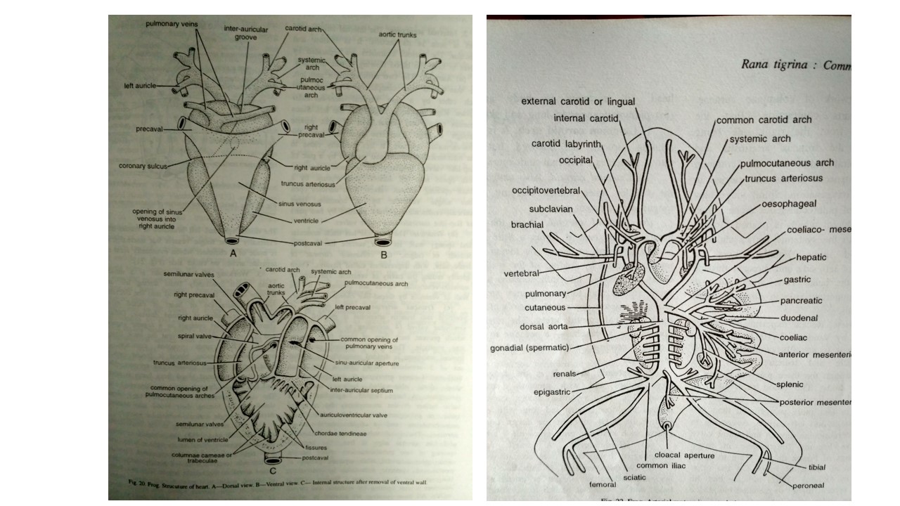
Arterial system of frog
Aortic arches:
- Arteries carry blood away from the heart.
- The initiation of arterial system starts with truncus arteriosus.
- The truncus divides into left and right branches and each of these branches subdivide into three major vessels or aortic arches:
- Common carotid to head
- Systemic to body and viscera
- Pulmocutaneous to lung and skin
i. Common carotid arch:
- It is a short vessel that runs forward and outward.
- It divides into 2 branches i.e. external and internal carotids.
- External carotid:
- It is also called lingual.
- It is the smaller inner branch that carries blood to the tongue and adjacent parts.
- Internal carotid:
- It is the larger outer branch.
- It forms a little swelling at its base, called as the carotid labyrinth or gland.
- Its lumen is converted into a labyrinth by folding of walls.
- A network of small vessels forms the inner cavity of carotid labyrinth which forms spongy structure.
- Probably it is a sense organ.
- It controls blood pressure in the internal carotid artery.
- The internal carotid artery divides into 3 branches:
- A palatine to the roof of mouth.
- A cerebral to the brain
- An ophthalmic to the eye
ii. Systemic arch:
- It is the longest of the three arches.
- It has the greatest distribution of blood.
- The two systemic arches curve dorsally around the oesophagus and join with each other behind the heart to form the dorsal aorta.
- Each systemic arch gives off 3 arteries:
- Oesophageal: It is a small artery to oesophagus. It may arise from occipito-vertebral.
- Occipito-vertebral: It immediately sends an occipital branch to occiput or posterior part of head, and a vertebral branch to vertebral column and spinal cord.
- Subclavian: It is a large artery that supplies the shoulder region and extends into the forelimb as brachial artery.
- All these arteries exhibit bilateral symmetry.
- However, from the left systemic arch, an additional branch is given off, the oesophageal which is absent in right side.
- Dorsal aorta:
- Dorsal aorta is formed by the union of both the systemic arches.
- It runs posteriorly lying mid-dorsally, just beneath the vertebral column.
- It gives off the following branches:
- Coeliaco-mesentric:
- It is a single layer artery.
- It arises from the junction of the two systemic arches.
- It has two main branches:
- Coeliac: to stomach, pancreas and liver
- Anterior mesenteric: to spleen and intestine
- Gonadial:
- It is a pair of short arteries to gonads.
- It is termed as spermatic in male frog and ovarian in female frog.
- Renal:
- While passing between the two kidneys, dorsal aorta sends off 5-6 pairs of small renal arteries in a series into both the kidneys.
- Posterior mesenteric:
- It arises from the posterior end of dorsal aorta, or sometimes from the anterior mesenteric.
- It goes to large intestine or rectum.
- Common iliacs:
- The dorsal aorta finally bifurcates posteriorly into two common iliacs.
- Each iliac supplies an epigastric to ventral body wall, recto-vesicular to rectum and urinary bladder, femoral to hip and upper thigh, and sciatic to lower leg.
iii. Pulmocutaneous arch:
- It divides into two main arteries:
- Pulmonary to the lung
- cutaneous to skin of dorsal and lateral sides and various other structures.
- Of these auricularis supply blood to tympanum, thymus gland, lower jaw, pharynx, and hyoid etc.
- However, dorsalis supplies the skin of dorsal side and lateralis supplies the skin of lateral sides of the body.
Venous system of frog:
- The venous system includes veins or those blood vessels in which blood of the body returns to the heart.
- In frog, it can be studied in 4 parts:
- Pulmonary veins
- Caval veins
- Renal portal veins
- Hepatic portal veins
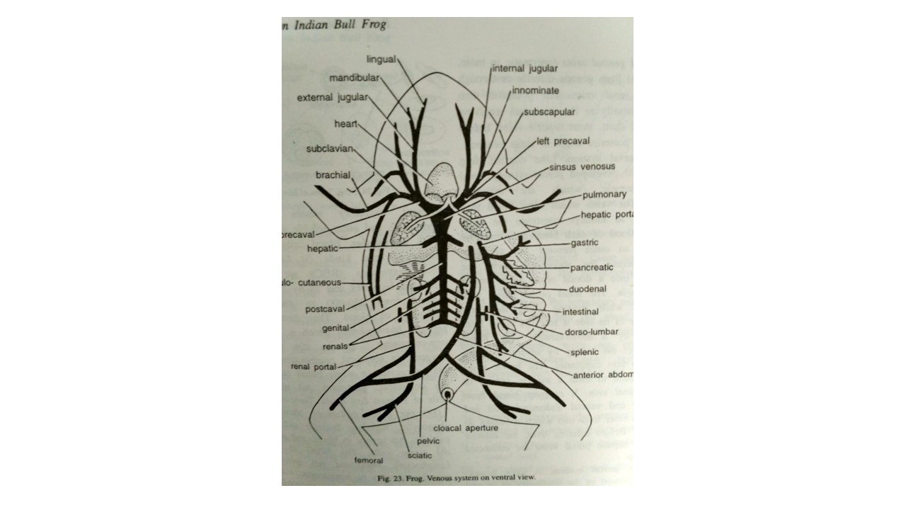
i. Pulmonary veins:
- The right and left pulmonary veins collects oxygenated blood from two lungs.
- These veins unite to form a common pulmonary vein opening directly into the left auricle on the dorsal side.
ii. Caval veins:
- Deoxygenated blood from rest of the body travels towards the heart in three large vessels, two anterior venae cavae and single posterior vena cava, all the three opening into sinus venosus.
- Anterior venae cavae or precavals:
- The right and left precavals or anterior venae cavae collect venous blood from the anterior part of body.
- Each precaval is formed by the union of 3 major veins which meet simultaneously:
- External jugular:
- It is formed by the slender and sinuous lingual from tongue and mandibular from outer margin of lower jaw.
- Innominate:
- It is formed by internal jugular from cranial cavity and orbit and subscapular from shoulder and back of arm.
- Subclavian:
- It is formed by the brachial from forelimb and the musculo-cutaneous from side of body and head.
- Posterior vena cava or postcaval:
- The single postcaval is a large, dark colored vein.
- It lies ventral to dorsal aorta.
- Its posterior end is formed between the two kidneys from which it drains blood by 5-6 pairs of renal veins.
- It also receives a pair of genital veins (spermatic in male, ovarian in female) from gonads directly or through anterior pair of renal veins.
- Then, the postcaval runs forwards, dorsally to the liver.
- It receives a pair of short, stout hepatic veins, before opening into the posterior angle of sinus venosus.
iii. Renal portal system:
- Frog has well developed portal systems:
- renal portal
- hepatic
- These portal systems are curiously interconnected.
- The veins which carry blood to a capillary system in kidneys forms the renal portal system.
- Blood of each hindleg is collected by two veins:
- an outer femoral
- an inner sciatic.
- On entering the abdominal cavity, the femoral divides into a dorsal renal portal and a ventral pelvic vein.
- The renal portal unites with the sciatic.
- While running along the outer border of kidney of its side, it receives blood from the lumbar region by a dorso-lumbar vein.
- Renal portal vein enters the kidney by various branches which break up into capillaries.
- The pelvic veins of both sides are united to form a median ventral or anterior abdominal vein.
- It receives blood from urinary bladder and ventral abdominal wall.
- Then, it runs towards to enter liver into which it breaks up into capillaries.
- The anterior abdominal and hepatic portal veins are connected by a small loop before entering liver.
iv. Hepatic portal system:
- A large hepatic portal vein is formed by the joining of several branches from stomach, intestine, spleen and pancreas.
- It carries blood of alimentary canal, heavily loaded with digested food stuffs, to the liver into which it breaks up into capillaries.
- The anterior abdominal vein is connected with hepatic portal vein, in the region of liver.
Blood of frog:
- Blood is the chief circulatory fluid of the body.
- It is actually a liquid connective tissue.
- It contains a clear liquid called plasma.
- In plasma, various types of free cells, called blood corpuscles are suspended.
- Plasma:
- Plasma forms nearly 2/3rd of the blood.
- It is largely water (90%) in which mineral salts, absorbed foods (sugars, proteins), excretory wastes (urea), secretions (hormones), and other soluble substances are dissolved.
- Corpuscles:
- Blood cells or corpuscles are mainly of 3 kinds:
- Erythrocytes or red blood corpuscles (RBC):
- These are oval, flattened, nucleated, biconvex and 14 by 23 micrometer in size.
- They number from 250,000 to 450,000 per cubic millimeter of blood.
- They bear the respiratory pigment haemoglobin.
- Hemoglobin is a yellow to red iron-containing protein.
- Hemoglobin serves to carry oxygen in chemical combination to tissues.
- Leucocytes or white blood corpuscles (WBC):
- They are colorless, nucleated and mostly amoeboid cells.
- It is of at least five types.
- They average 5,000 to 7,000 per cubic millimetre.
- Most of them are phagocytic, ingesting bacteria and other foreign particles that appear in blood.
- They also remove dead or old tissue cells.
- The WBC of frog are: lymphocytes, monocytes, and granulocytes.
- The granulocytes may be neutrophilic, eosinophilic and basophilic.
- Thrombocytes or blood platelets:
- These are small, nucleated, spindle cells.
- They play an important role in coagulation.
- When a blood vessel is injured, the disintegration of thrombocytes releases an enzyme thrombin.
- It changes soluble fibrinogen of blood plasma into insoluble fibrin.
- The latter forms the clot which seals the wound to prevent further loss of blood.
- Blood cells are produced mainly in bone marrow and spleen, the latter also destroys the worn-out cells.
- Lymphatic system:
- Lymphatic system of frog is more primitive than that of higher vertebrates.
- It is made of lymph, lymph vessels, lymph hearts, lymph spaces and spleen.
Lymphatic system of Frog
- Lymph:
- Blood confined to blood vessels does not come directly in contact with body cells and tissues.
- It is filtered regularly from capillaries into intercellular spaces forming the tissue fluid or lymph.
- Lymph bathes the tissues and lubricates many of the internal organs.
- Being a filtrate of blood, it closely resembles plasma containing leucocytes.
- However, it lacks erythrocytes and some blood proteins.
- It functions as the middle man passing over food and oxygen to cells and waste materials to blood.
- The tissue fluid is continually being removed into lymph vessels in which it is called lymph.
- Lymph vessels:
- Lymph vessels or lymphatics are thin-walled delicate vessels of various sizes, forming networks but are difficult to see.
- Lymph capillaries unite to form larger vessels which open into the venous system, thus returning lymph back to blood from which it originates.
- Some small openings in the peritoneum communicate with lymph vessels.
- Lymph hearts:
- Lymph vessel open into veins at four places in frog.
- At each opening, the lymph vessel forms, called a lymph heart.
- Lymph heart is a small rhythmically contractile sac which slowly drives lymph into vein.
- Thus, frog has two pairs of lymph hearts.
- One pair is situated anteriorly below the scapulae that opens into subscapular veins.
- Another pair lying posteriorly on either side of the tip of urostyle open into femoral veins.
- Lymph spaces:
- Unlike other vertebrates, in frogs and toads, some lymph vessels become dilated to form huge lymph channels or sinuses.
- There are extensive subcutaneous spaces under skin (dorsal, lateral, abdominal etc.) due to which the skin is loosely attached.
- These spaces are separated by connective tissue septa.
- One of the most important is the sub-vertebral lymph space above kidneys
- Spleen:
- The spleen is a small, round, and dark red gland.
- It is located in the mesentery near rectum.
- It contains the largest mass of lymphatic tissues in body.
- It destroys worn out erythrocytes and produces antibodies, new erythrocytes and lymphocytes which are phagocytic in nature.