Mode of transmission:
- Infected person with circulating microfilariae is the chief source and reservoirs of infection.
- Person to Person transmission occurs by the bite of infected mosquitos.
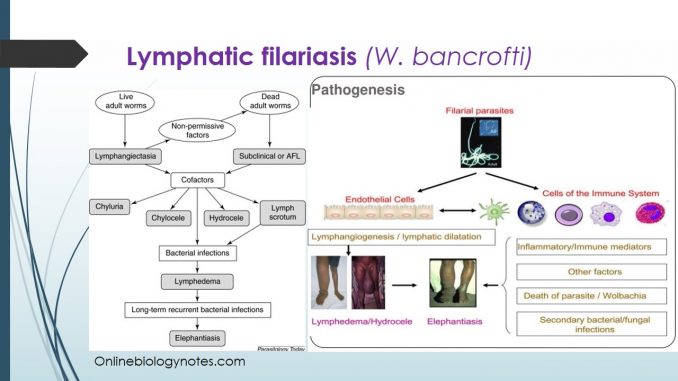
Pathogenesis:
- Though L3 larva are infective larva they do not have any pathogenic effect.
- Similarly, circulating microfilariae are also not pathogenic.
- The pathogenic effects are due to the fourth stage larva, moulting stage from L3 and adult worms whether living or dead present in the lymphatic vessels.
- The following stage occur sequentially during the pathogenesis or lymphatic filariasis.
- i. Dilation of lymph vessels:
- The developing larva and metabolic products released during the moulting of the larva, unsheathing of microfilariae during moulting and the presence of the adult worms induces inflammatory reactions.
- The intense inflammation of lymph vessels leads to dilation of lymph vessels during the early stage of infection.
- Besides inflammation, immune reaction of host against the worms and toxic effects of the worms also results in dilation of lymphatic vessels.
- Dilation of lymphatic causes an increased secretion of proteinaceous material from lymphatics into the surrounding tissue leading to the information of conspicuous lymphedema and thickening of the endothelium.
- ii. Infection of lymphatic vessels (lymphangitis):
- Progression of infection develops with lymphangitis.
- It is characterized by presence of dilated, inflamed and thickened lymphatic vessels associated with erythema, edema and tender painful areas.
- The main causes of lymphangitis are:
- Irritation caused by the movement of the adult worm inside the lymphatic system.
- Release of metabolic wastes by larva
- Absorption of toxic wastes liberated from dead worms by host cells.
- Secondary bacterial infection streptococci.
- iii. Obstructions of the lymph node:
- The lymphangitis is followed by necrosis, sclerosis and obstruction of lymphatic vessels proximal to the lymph nodes.
- Flow of lymph is obstructed due to-
- Presence of worm in the lymph vessels
- Thickening to lymphatic vessels as well as focal necrosis.
- Giant cell formation, fibrosis as well as cellular changes result in obstruction of lymphatic vessels
- The obstruction of the lymph flow results in elephantiasis which is the classical feature.
- The course of the events in the pathogenesis of the lymphatic filariasis are variable and depend upon interaction of a variety of host and parasitic factors.
Clinical manifestation: Lymphatic filariasis
- It is caused by the juvenile and adult worms of W. bancrofti.
- The clinical manifestation of the condition depend on stages of the disease as follows:
i. Endemic normal: No overt clinical symptoms
ii. Asymptomatic stage:
- Person in this stage have microfilariae in their blood but do not show any clinical manifestation of filariasis.
- They may remain asymptomatic for years or even after life.
iii. Acute filariasis:
- Acute filariasis or inflammatory phase is caused by antigens released from female adult worms.
- The condition is characterized by Filarial fever (usually low grade but occasionally severe), accompanied by chills, general malaise, headache and pain are other symptoms.
- Lymphedema
- Lymphadenitis
- Adeno-lymphangitis (ADL)
iv. Chronic filariasis:
- It is the obstructive phase usually takes 10-15 years to develop.
- Typical manifestation includes:
- Lymph varices: Caused by the obstruction of lymph flow and accumulation of lymph in the ducts leading to dilation of the ducts.
- Hydrocele: Caused by obstruction of the lymph vessels of the spermatic cord and exudation from the inflamed test and epididymis.
- Elephantiasis: It is the result of wuchererial infection and usually follows years of continual infections.
- It is caused by fibrotic construction of all the afferent lymphatics draining the past.
- Hypertrophy and hyperplasia seen are the result of excessive protein, in the lymph exudates stimulating the connective tissue to excessive growth.
- Elephantiasis in the scrotum, legs and arms of male and legs and arms of female is the feature of chronic elephantiasis.
- The affected part becomes enormously enlarged producing a tumor like solidity.
- The surface of the skin becomes rough, and even papillomatous.
- The hairs become rough and sparse.
- On section the skin cuts like an unripe pear, it is thickened, dense and fibrous.
- The subcutaneous tissue shows a blubbery appearance in which the dilated and thickened lymphatics and veins can be seen.
- The underlying muscles and bones do not usually show any alteration.
- Granuloma of breast: Characterized by the presence of film solitary mass in the breast.
- Chyluria: Urine shows chyle mixed with blood and occasionally microfilariae.
- Caused by escape of chyle through the urine due to the rupture of varicose chyle vessels through the mucous membrane of the urinary tract.
v. Occult filariasis:
- It denotes a condition of hypersensitivity reaction of the host to micro-filarial antigens characteristically microfilariae are not found in the peripheral blood and the classic features of lymphatic filariasis are absent.
- Tropical pulmonary eosinophilia (TPE) is the most important manifestation.
- Arthritis, tenosynovitis, dermatoses etc. in the endemic areas are the less frequent manifestation of occult filariasis.
- TPE is distinct clinical syndrome characterized by chronic pulmonary infiltration in chest X-ray, hypereosinophilia of the peripheral blood and respiratory symptoms like low grade fever, cough, chest pain and asthmatic attacks especially at night.
vi. Less frequent lesions:
- These includes granuloma of the spleen and other organs and the presence of adult W. bancrofti in the anterior chamber of the eye.
Complications:
- Secondary bacterial infections of the overlying skin of elephantiasis of the leg or arm.
Laboratory diagnosis:
Parasitic diagnosis:
- Specimen: Peripheral blood is the specimen of choice.
Methods of examination includes:
i. Microscopy:
- The standard method for diagnosing active infection is the identification of microfilariae in blood smear by microscopic examination.
- Blood collection should be done at night to coincide with the appearance of the microfilariae.
- It can be determined by following method:
- Direct wet mount:
- 2-3 drops of blood are collected on a clean glass slide and examined microscopically after placing cover slip on it.
- -ive microfilariae are identified by their characteristic serpentine movement in the blood plasma.
- Stained thick blood film smears:
- Thick and smear stained with Giemsa or Leishman is the most commonly used method.
- The presence of sheath but the absence of nuclei in the tail end of microfilaria is diagnostic of W. bancrofti microfilaria.
- Concentration of blood:
- Increase of low number of microfilariae in blood, their recovery can be increased by various concentration methods like knot’s method of concentration by sedimentation, membrane filtration concentration methods using Nucleopore or Millipore membrane filters.
- DEC provocation test:
- In this test (Diethylcarbamazine) is given orally at a dose of 2-8 mg/kg. after 30 min the capillary blood is collected by finger prick for demonstration of microfilariae by direct wet amount or staining the smear.
- DEC stimulates nocturnal periodic microfilariae to circulate in the peripheral blood during the day time.
- QBC:
- This method can frequently demonstrate the circulating microfilariae in the blood.
- Urine microscopy:
- Microfilariae can be demonstrated in the chylous urine.
- 10 ml-20 ml of the first early morning urine is collected for examination and demonstration of microfilariae by microscopy.
- Microscopy of hydrocele fluid and lymph node aspirations.
- Microfilariae can also be demonstrated in hydrocele fluid and also in lymph node aspiration. Either is used hydrocele fluid to dissolve fat globules.
II. Immune diagnosis:
Serological tests:
i. Demonstration of circulating antibodies:
- IHA, IFA, ELISA, RLA, luminescence immune analyses are used to demonstrate the circulating antibodies in the serum
- Disadvantage of these test are that they show cross reactivity with sera from other filarial and helminthic infections and they are unable discriminate between past and current infections.
ii. Demonstration of circulating antigens:
- The circulating antigens are present in serum only during recent or current infections.
- ELISA employing monoclonal antibody AD12 detects a 200 Kda of adult W. bancrofti in the serum.
- The other ELISA using monoclonal antibiotic Og4c3 detects adult worm as well as microfilariae antigen in the serum.
Molecular methods:
- PCR methods have been developed however they are not much sensitivity PCR is positive only when circulating microfilariae are found in the peripheral blood.
Imaging methods:
- X-ray: Chest x-ray shows diffuse pulmonary infiltrates in patients with TPE.
- Ultrasound: Only non-invasive method for detection of adult worms in the affected lymph nodes.
- The live adult worms are identified by a distinctive pattern of their movement known as filarial dance sign.
Other tests:
- Biopsy of lymph node that has been enlarged show cross sections of adult worms
- Eosinophilia can be seen in complete blood cell count
Treatment:
- Diethylcarbamazine (DEC), drug of choice.
- Dose: oral, 3mg to 6 mg/kg daily in divided doses for 3 weeks
- Others: Ivermectin, Levamisole, Mebendazole and Centprazine.
Prevention and control:
- Clinical control of mosquito by spraying DDT, malathion etc.
- Biological control by using of B. sphaericus, B. thuringienesis, Poecilid reticulate molliensis.
- Effective drainage and sewage system to eliminate breeding of mosquito
- Treatment of cases
- Use of bed nets, house screens
- Interrupting transmission of infection.
