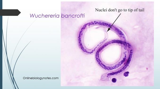
- Wuchereria bancrofti or Bancroft filarial worm is a parasitic filarial nematode spread by a mosquito vector.
- It is one of the three parasites that causes lymphatic filariasis (commonly known as elephantiasis), an infection of the lymphatic system by filarial worms.
- The parasite as named after physician Otto Wucherer and parasitologist Joseph Bancroft both of whom extensively studied the filarial infections.
Habitat
- Adult worms are found in the lymphatic vessel, especially the lymph nodes.
- The microfilariae are found in the peripheral blood, occasionally they are also found in chylous urine or in hydrocele fluid.
Morphology of W. bancrofti
1. Adult worms
- W. bancrofti exhibits considerable sexual dimorphism.
- These are minute, long hair like transparent (often creamy in color) nematodes.
- They are filiform in shape with both ends tapering.
- The head end terminating in a slightly round swelling, and surrounded by two rows of 10 sessile papillae. The posterior end contains anus at its terminal end.
- The male measures 2.5-4 cm in length with 0.1 mm in thickness. The tail end is curved ventrally and contains two spicules of unequal length.
- The females are longer than males measure 8-10 cm in length with 0.2-0.3 mm in thickness. Its tail end is narrow and abruptly pointed. The females are oviparous.
- The adults obtain their nourishment from the lymph of the lymphatic system.
- The life span of the adult worms is long, probably several years (5-10 year or even more).
2. Microfilariae (Embryos):
- The first stage of larva is called microfilariae.
- They are very active in their habits and can move both with and against the blood stream, when sustained, they appear as colorless and transparent bodies with blunt heads and pointed tails.
- The embryo measures about 290 mm in length by 6-7 mm in breadth.
- When dead and stained with Romanowsky’s stains, they show the following morphological features:
- Hyaline sheath:
- It is a sac like envelope which is much longer (359 mm) than the larval body represents the chorionic envelop of the eggs.
- It remains as investing membrane around the larva.
- Cuticle:
- It is lined by subcuticular cells and is seen only with vital stains.
- Somatic cells or nuclei:
- Nucleiappear as granules in the central axis of the body and extend from the head to the tail end, except the terminal 5% of the tip of the tail. This is the distinguishing feature of the parasite.
- The space at the anterior end devoid of granules is seen called as cephalic space.
- The granules are broken at definite places serving as the landmarks for identification of the species.
- They include:
(a) Nerve ring; an oblique space
(b) anterior V-spot represents the rudimentary excretory system
(c) the posterior V spot or tail spot represents the terminal part of the alimentary canal/anus or cloaca.
3. Third stage of larva (infective form):
- The L3 larva the infective form of the parasite is found only in mosquito.
- They are elongated, filariform, measures 1.4-2 cm in length and 18-23 cm in breadth.
Life cycle:
- W. bancrofti completes its life cycle in two hosts:
- Definite host: Human
- Intermediate host: mosquito, belonging to genus Culex, Aedes and Anopheles.
- Life cycle in Human: Entrance in the human and development into adult worms
- Infection is acquired by the bite of infected mosquito during which L3 larva are deposited on the skin.
- The L3 larva are not directly injected into the blood stream.
- The L3 larva are deposited on the skin near the site of the puncture.
- Later attracted by the warmth of the skin, the larva enters through the puncture wound or penetrates through the skin on their own.
- The L3 larva after penetrating the skin, reaches the lymphatic channels, settles down at some spot (inguinal, scrotal or abdominal lymphatics), metamorphose and becomes sexually mature.
- The male fertilizes the female and the gravid females discharge microfilariae which usually appear in the peripheral blood in 8-12 month of infection.
- These micro filariae circulate in the blood for 6 months to 2 years and then die if not taken by mosquito.
- Life cycle in Mosquito: Stages in the development of micro filaria
- Microfilaria ingested by the mosquito lose their sheath within 2 to 6 hours of their arrival in the stomach.
- Then they penetrate the gut wall and migrate to the thoracic muscle, where they rest and begin to grow.
- In the next 2 days, microfilaria become thick, short sausage shaped with a short spiky tail, measuring 124-200 mm in length 10-17 mm in breadth. This is the first stage larva L1.
- The larvae possesses a rudimentary digestive tract.
- During 3-7 days of time, the larva grows rapidly, moults once or twice and measures 225-330 mm in length by 15-30 mm in breadth. This is the second stage larva L2.
- Metamorphosis completes by 10-11days with distinct features such as the tail atrophies to a mere stump and the digestive system, body cavity and genital organs are now fully developed. This is the third stage larva L3.
- These L3 larva are the infective form which enters the proboscis sheath of the mosquito on or about the 14th day.
- When the mosquito bites a man during the blood meal, the L3 larva are released from the tip of proboscis of mosquito and the cycle is repeated.
- Development in mosquito takes place within 10-20 days.
Epidemiology of Wuchereria bancrofti
- W. bancrofti is largely confined to tropics and subtropics. They are found in India, West-Indies, Puerto Rico, Southern China, Japan, Pacific Island, West and central Africa, South America.
- The disease is endemic in 83 countries with more than 1.2 billion at risk.
- It is estimated that more than 120 million people are infected.
- More than 25 million men suffer from genital symptoms and more than 15 million people suffer from lymphedema or elephantiasis of leg.
Periodicity:
- The microfilariae of oriental countries (India and China) show nocturnal periodicity.
- They are found periodically in peripheral blood at night especially between 10pm to 4 am.
