
Trichuris trichiura
- Trichuris trichiura commonly called the whipworm because of its characteristic whip-like shape.
- It causes trichosis in human which is an intestinal infection caused by invasion of the colon by the adult worm.
Habitat:
- The adult worm lives in the large intestine (caecum) of human
- It can also be present in the vermiform appendix and rectum.
Morphology
- The adult worms resemble the whip. The anterior 3/5th of the end is very thin hair like and the posterior 2/5th is thick and stout resembling the handle of a whip.
- The anterior ends penetrate the mucosa layer and remain deeply embedded.
- Adult worms are pinkish in color.
- The anterior end consists of a long oesophagus which is a minute channel while the posterior end contains the intestine and sex organs.
Male worm:
- The adult males are 3-4 cm in length and are recognized by their characteristic coiled posterior end.
Female worm:
- Female worms are longer than males measuring 4-5 cm in length.
- The caudal extremity is either comma shaped or an arc shaped
Eggs
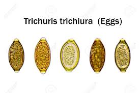
- Egg of Trichuris trichiura has diagnostic value. Egg is barrel shaped with a mucous plug at each end.
- It is brown colored (bile-stained) and has a double shell.
- The egg measures 50-54 µm in length and 22-23 µm in breadth.
- Eggs contain an un-segmented ovum when it leaves the human hosts.
- The freshly passed eggs are non-infective to human.
- Eggs float in saturated NaCl solution.
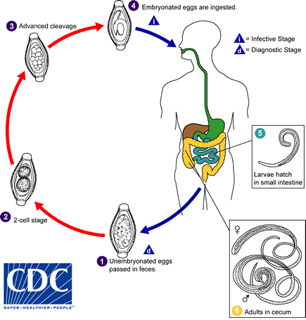
Life cycle of Trichuris trichiura:
- The life cycle of T. trichiura is simple and is completed in single host, the man. However, change of host is needed for the continuation of species.
- No intermediate host is required.
- Human acquires infection by ingestion of food or water contaminated with embryonated eggs.
- The digestive enzymes dissolve the eggs shell and the larva emerges out through one of the poles of the eggs.
- The liberated larva then pass down into the caecum which is their site of the localization.
- In caecum the larvae develops into adult worm and become sexually mature within a month from the time of ingestion of eggs.
- The female worm after being fertilized by the male begins to lay the eggs, which is about 3 month after infection.
- The freshly laid eggs are un-embryonated and excreted out with the faeces.
- Each adult female can produce about 5000-7000 eggs per day for upto 5 years.
- Embryogenesis within eggs occur in outside environment in the water or damp soil.
- In tropical climates larva develops within the egg in the course of 3-4 weeks. In temperate climates, the larva takes a long time (6-12 month) to complete its development.
- Once the egg is embryonated, it is infective to human.
Mode of transmission:
- The food, water and soil contaminated with embryonated eggs are the chief sources of infection.
- Ingestion of embroynated eggs in the contaminated food and water
- Contaminated fingers during soil works
Pathogenesis
- The adult worm invades the intestinal mucosa by its thin, thread like anterior end and feeds on tissue secretions but not on blood.
- It causes petechial hemorrhage, inflammation, oedema and mucosal bleeding in the intestinal mucosa at their site of attachment.
- The lumen of appendix can be blocked in case of severe worm load.
- Presence of worms in the mucus membrane irritates the nervous plexus of mucosa causing diarrhea and cramps.
- Occasional eosinophilia can be present
- Approximately 0.005 ml of blood per worm per day is lost in the infected man.
Clinical manifestation
- The clinical manifestation of Trichuris trichiura depends upon the intestinal worm load of the person.
- Infection is asymptomatic in case of light infection with 100-200 worms.
- For moderate infection the number of worm should be more than 200 worms and this can manifest as vague abdominal discomfort and diarrhea (rarely bloody), vomiting, headache etc.
- Trichuriasis: In case of heavy infection with more than 800 worms, serious complications especially in children are observed.
- It causes bloody diarrhea with profuse mucus, abdominal pain and tenesmus weight loss leading to the cachexia, severe anaemia.
- Distribution of a large number of worms throughout the colon and rectum may cause prolapse of the rectum.
- Migrating worms can occasionally cause appendicitis.
Epidemiology
- About 800 million people are affected worldwide
- Trichuris trichiura infection is commonly found in Tropical and subtropical countries with moist and warm soil. Such as tropical Africa, South America and South East Asia.
Diagnosis
- Specimen: stool, blood
- Microscopy:
- Finding of characteristic barrel-shaped eggs in the faeces on light microscopy.
- Stool concentration methods may be required to detect light infection
- Adult worms may occasionally be present in the stool.
- The degree of infection can be determined by egg count.
- In heavy infection stool is frequently mucoid and contains charcot-Leyden crystals.
- Proctoscopy: adult worm can be obtained from rectal mucosa sample.
- Blood test: shows eosinophilia
Treatment
- Mebendazole- drug of choice
Dose: 100 mg twice daily for 3 days
- Albendazole – 400 mg daily for 3 days
Prevention and control
- Sanitary disposal of faeces.
- Personal hygiene
- Food hygiene
- Treatment of infected cases.