
Anatomical structure of Monocot root:
T.S. of monocot root shows the following anatomical features:
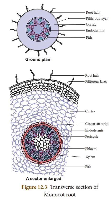
Epidermis/Epiblema/Rhizodermis:
- It is the outermost layer composed of compact parenchymatous cells having no intercellular spaces and stomata.
- The tubular unicellular root hairs are also present on this layer
- Both epiblema and root hairs are without cuticle.
- In older parts, epiblema either becomes impervious or is shed.
- Epiblema and root hairs absorb water and mineral salts.
Cortex:
- It lies just below the epidermis.
- Cortex consists of thin walled multilayered parenchyma cells having sufficiently developed intercellular spaces among them.
- Usually in an old root of Zea mays, a few layers of cortex undergo suberization and give rise to a single or multi-layered zone- the exodermis.
- This is a protective layer which protects internal tissues from outer injurious agencies.
- The starch grains are abundantly present in the cortical cells.
- Cortex functions as:
- a) conduction of water and mineral salts from root hairs to inner tissues
- b) storage of food
- c) protection when exodermis is formed in older parts.
Endodermis:
- The innermost layer of the cortex is termed as endodermis.
- It is composed of barrel-shaped compact cells that lacks intercellular spaces among them.
- Young endodermal cells have an internal strip of suberin and lignin which is called casparian strip.
- The strip is located close to the inner tangential wall.
- There are some unthickened cells opposite to the protoxylem vessels known as passage cells which serve for conducting of fluids.
- The function of endodermis is to regulate the flow of both inward as well as outward.
Pericycle:
- It lies just below the endodermis and is composed of single layered sclerenchymatous cells intermixed with parenchyma.
Vascular tissue:
- The vascular tissue contains alternating strands of xylem and phloem.
- The phloem is visualized in the form of strands near the periphery of the vascular cylinder, beneath the pericycle.
- The xylem forms discrete strands, alternating with phloem strands.
- The center is occupied by large pith which maybe parenchymatous or sclerenchymatous.
- The number of vascular bundles is more than six, hence called as polyarch.
- Xylem is exarch i.e. the protoxylem is located towards the periphery and the metaxylem towards the center.
- Vessels of protoxylem are narrow and the walls possess annular and spiral thickenings in contrast, metaxylem are broad and the walls have reticulate and pitted thickenings.
- Phloem strands consist of sieve tubes, companion cells and phloem parenchyma.
- The phloem strands are also exarch having protophloem towards the periphery and metaphloem towards the center.
Conjunctive tissues:
- In between the xylem and phloem bundles, there is the presence of many layered parenchymatous or sclerenchymatous tissue.
- These help in storage of food and help in mechanical support.
Pith:
- It is the central portion usually composed of thin-walled parenchymatous cells which appear polygonal or rounded in T.S.
- Intercellular spaces may or may not be present amongst pith cells.
- In some cases pith becomes thick walled and lignified.
- Pith cells serve to store food.