
Anatomical structure of dicot stem
T.S. of dicot stem shows following internal features:
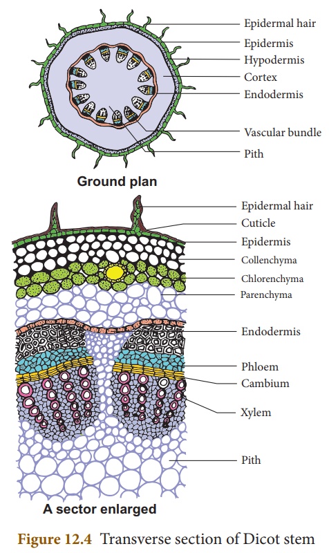
Epidermis:
- It is the outermost layer and has a single layer of parenchymatous cells.
- It possesses stomata and large number of multicellular hairs (trichomes).
- The outer walls are greatly thickened and cutinized.
- The cells are compactly arranged and do not possess intercellular space.
- The epidermis has following functions:
- Minimize the rate of transpiration owing thick cuticle
- Protects the underlying tissues from mechanical injury
- Prevents the entry of harmful organisms
- Helps in the exchange of gases through stomata.
Hypodermis:
- This layer lies below the epidermis and is composed of 4 or 5 layers of collenchymatous cells.
- These cells are specially thickened at the corners against the intercellular spaces due to deposition of cellulose and pectin.
- The cells are living in nature and may contain few chloroplasts.
- It provides mechanical strength and elasticity to the peripheral portion of the stem particularly the young and growing organs.
- They perform photosynthesis and also acts as storage of food.
Cortex:
- It lies below the hypodermis.
- It consists of a few layers of thin-walled, large, rounded, or oval, living parenchymatous cells, having intercellular spaces.
- Cells of cortex may contain some chloroplasts which may function to manufacture of food materials.
- They serve for storage of food.
Endodermis:
- It is the single innermost layer of the cortex which separates the cortex from vascular bundles.
- Cells are somewhat barrel shaped and compactly arranged, having no intercellular spaces and are parenchymatous.
- Usually, the cells contain starch grains and thus the endodermis maybe termed as starch sheath.
- They serve as food reserve.
- The radial and the transverse walls are thickened due to the deposition of lignin forming casparian strips.
Pericycle:
- It lies in between the endodermis and vascular bundles.
- It is generally composed of sclerenchymatous and parenchymatous cells.
- The sclerenchyma is in the form of semilunar patches above the vascular bundles which give mechanical support to the plant parts.
- Similarly, parenchymatous pericycle is present outside the medullary rays which serves to store food.
Vascular bundles:
- These are arranged in a ring around the central pith and inner to the pericycle.
- These are conjoint, collateral, open and wedge-shaped.
- The size of the bundles varies in different species.
- Each bundle has a patch of xylem towards the center, a patch of phloem towards the periphery and a strip of cambium in between them.
- Xylem:
- It lies towards the pith of vascular bundles.
- It consists of tracheids, vessels, xylem parenchyma, xylem fibers.
- Tracheids and vessels consists of smaller protoxylem and larger metaxylem.
- Protoxylem is first formed that lies towards the center but metaxylem is later formed that lies towards the periphery.
- This type of xylem is called endarch xylem. It helps in conduction of sap.
- Phloem:
- It lies just below the sclerenchymatous patch of pericycle and is composed of following elements such as sieve tubes, companion cells, and phloem parenchyma.
- It conducts the foods.
- Cambium:
- It lies in between xylem and phloem.
- It consists of a narrow strip of meristematic cells having large nuclei and dense cytoplasm, called fascicular cambium.
- It is responsible for secondary growth in thickness of the plant body.
Pith:
- It occupies the central portion of the stem.
- It is composed of thin walled parenchymatous cells which are rounded or polygonal, with or without intercellular spaces.
- Food is stored in this region.
- Medullary rays:
- These are the thin-walled, radially elongated parenchymatous cells present in between vascular bundles.
- These store food materials and help in internal translocation of water.