What is Immunoassay?
- An immunoassay is a biochemical test that measures the concentration of a substance in a liquid (a portion of a biological specimen) using the reaction of an antibody or antibodies to its antigen (drug).
- They are used in a lot of laboratories, including hospitals labs, and have been widely used in the special area of Forensic toxicology to screen for drugs and other chemicals in the body.
- Immunology is a laboratory science that studies the body’s immunity to disease.
- The basic mechanism of immunity is the binding of drugs or other chemical compounds to antibodies (large proteins produced by the body’s immune system).
Principle of Immunoassay:
- An assay is a general term for an analytical laboratory procedure designed to detect the presence of and/or the quantity of a drug in a biological fluid such as urine or serum (the fluid component of the blood obtained after removal of blood cells and fibrin clot).
- An immunoassay, therefore, is an analytical procedure which has as its basis the principles of immunology- specifically the binding of drugs to antibodies.
- This binding of antibodies to drugs forms the basis for immunoassay.
- In the development of an immunoassay, the first step is to inject an animal (host) with the drug that we ultimately wish to analyze.
- The host immune system, recognizing the drug as a ‘foreigner’, generates antibodies to this drug, and these antibodies can then be harvested from the serum of the animal.
- In the test-tube environment of the laboratory (in vitro), these antibodies can be recombined with the appropriate drug.
- Just as it did inside the body (in vivo), the antibody will recognize the drug based on the lock and key fit and will spontaneously bind to it.
- The second step in the development of an immunoassay is to synthesize a ‘labelled’ drug.
- This involves the chemical addition of a ‘marker’ to the drug.
- This marker can be small, such as an atom of radioactive iodine, or it can be large, such as an enzyme, which is a fairly large protein.
- Irrespective of its size, this marker is added on such a way that it doesn’t interfere with the lock-and-key recognition between the antibody and the drug.
- All immunoassays work in the same basic fashion.
- They differ in the types of labels that are added to the labeled drug and in the analytical methods by which the amount of binding of labeled drug to the antibody is measured.
Types of Immunoassay
- Several different types of immunoassay are routinely performed in the laboratory.
- Although they differ in the types of reagents and instrumentation used, they are all based on the same scientific principle (the binding of drugs to antibodies).
- The three types of immunoassay that are commonly used for drug testing are the radioimmunoassay (RIA), enzyme multiplied immunoassay (EMIT), and fluorescence polarization immunoassay (FPIA).
- The immunoassay is based on the competitive or non-competitive binding of the antigen with the antibody.
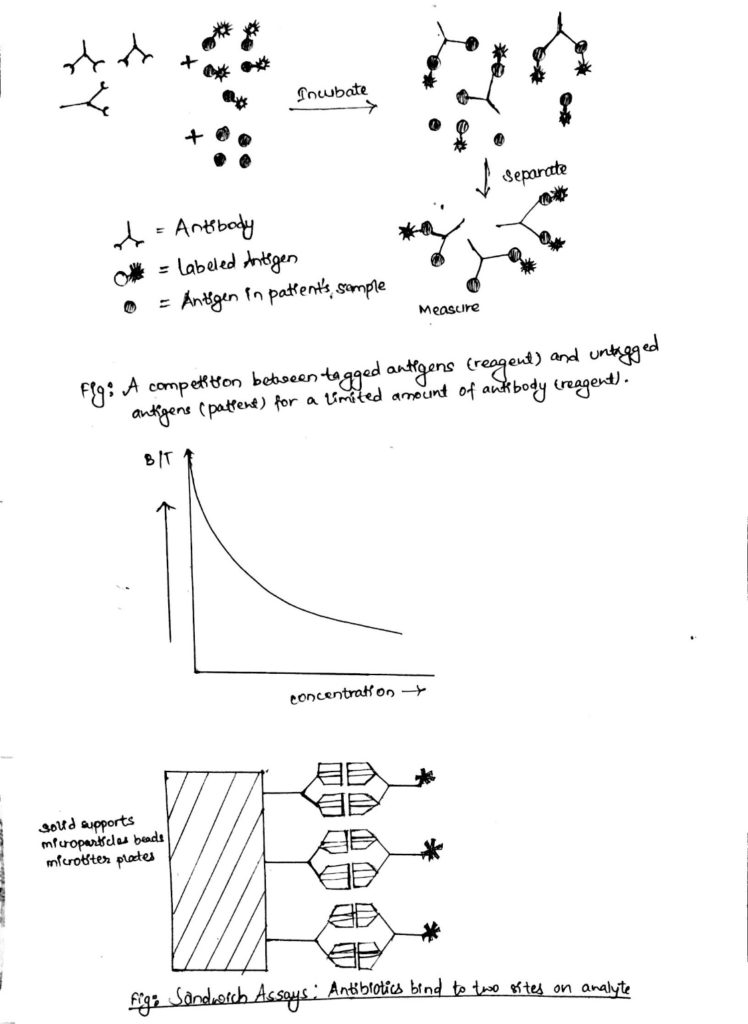
1. Competitive immunoassay
- (Competition between tagged and un-tagged antigen for the antibody)
- Competitive Immunoassays are always designed so that there are fewer antibody-binding sites present in the reaction mixture than there are molecules of (labeled plus unlabeled) drug.
- Because the label and unlabeled drug appear the same to the anti-body, they will compete equally for the limited number of available binding sites on the antibody.
- By measuring the amount of labeled drug bound to the antibody, the analyst can calculate the amount of unlabeled drug in the biological specimen.
- Commercially available immunoassay kits contain the antibody (which the company has prepared described above) and the labeled drug (which has been chemically synthesized) necessary to perform the assay.
- In the laboratory, a fixed amount of antibody and a fixed amount of labeled drug are placed into a reaction vessel (test tube).
- If these were the only two ingredients, all the binding sites on the antibody would react with (bind) to the labeled drug.
- A third ingredient added to the assay is, however the unlabeled drug (i.e., the urine, saliva, or serum specimen containing the drug that is being measured).
- Because the label on the labeled drug is placed in a position that doesn’t interfere with binding to the antibody (i.e., it is ‘hidden’), the antibody cannot distinguish between the labeled and unlabeled drug.
- Mix the three components together.
- Allow the antigens to compete for the limited antibody
- Antibody will bind with tagged or untagged antigen
- Separation step: antibody-antigen complexes are separated from free antigens
- Tagged antibody-antigen complex is measured.
- The tagged antigen and antibody from the reagent kit are constant.
- The only variable is the concentration of the patient antigen (the thing we want to measure)
- A standard curve can be made with known antigen concentrations giving the following general results:
- High concentrations of patient antigen mean that more of the antibody-antigen complexes are untagged
- Low concentrations of patient antigen mean that more of the antibody-antigen complexes are tagged
- There is an inverse relationship between patient antigen concentration and tag activity after the separation process.
- The activity of the tag is measured twice:
- Before separation step= Total tag activity
- After separation step = Bound tag activity (antibody-antigen complex)
- Note that the separation process removes all unbound (free) tag from the testing
- %B= B/T X 100
- The ratio of the bound activity to the Total activity (B/T) decreases as the concentration of the patient’s (untagged) antigen increases.
- Using standard solutions of known antigen concentrations, the %B is plotted against the concentrations of the standards.
2. Non-competitive immunoassays:
- Noncompetitive (sandwich) immunoassays generally provide the highest level of assay sensitivity and specificity.
- This format is referred to as a ‘sandwich’ assay because the analysis id bound (sandwiched) between two highly specific antibody reagents.
- The reaction mixture typically includes an excess of labeled antibody, so that all drug/metabolite is bound.
- The amount of antibody-antigen complex is then measured to determine the amount of drug present in the sample.
- The measurement of labeled analyte, usually antibody, is directly proportional to the amount of antigen present in the sample.
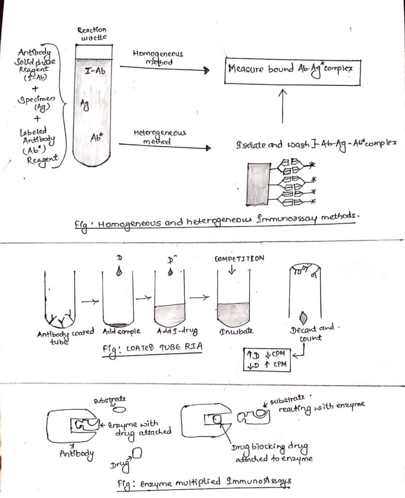
3. Homogeneous and Heterogeneous Immunoassay methods:
- Immunoassay methods that require separation of bound Ab-Ag* complex are referred to as heterogeneous immunoassays.
- Those that do not require separation are referred to as homogeneous immunoassays.
- Homogeneous methods have been generally applied to the measurement of small analytes such as abused and therapeutic drugs.
- As homogeneous methods do not need the separation of the bound Ab-Ag* from the free Ag*, they are generally much easier and rapid to perform.
Types of Immunoassays used for drug assay:
i. Radioimmunoassay:
- Radio-immunoassays (RIAs) utilize a radioactive label (usually 125I, 3H or 14C), which emits radiation that can be measured with a beta or gamma counter.
ii. Enzyme multiplied immunoassay:
- In the Enzyme multiplied Immunoassay (EMIT), the drug in the sample and the drug labeled with G6PD compete for antibody binding sites.
- Binding inhibits enzyme activity, while free enzyme remains active to interact with.
- Enzyme activity/absorbance is directly proportional to drug concentration.
iii. Fluorescence Polarization Immunoassay:
- In the Fluorescent Polarized Immunoassay, the drug in the sample competes with fluoresce labeled drug for antibody binding sites.
- Reaction mixture is excited by plane polarized light.
- As the tracer returns to a lower energy state, it emits light, polarization is measured.