
Ascaris lumbricoides: Morphology, life cycle, Pathogenesis, lab diagnosis and Treatment
Ascaris lumbricoides is an intestinal round worm. It is the largest intestinal nematode to infect Human. The adult worm lives in small intestine and grow to a length of more than 30 cm. Human is only the natural host and reservoir of infection.
The round worm infection occurs worldwide. The number of infected persons is estimated to be more than 2 billion. The main epidemic region with prevalence rate of approx. 10-90% includes countries on South east Asia, Africa and latin America.
Morphology:
Adult:
The round worm resembles to earthworm. It is elongated tapering to both end, anterior being thinner than posterior. Freshly excreted worm is yellowish pink in color, which gradually changes to white.
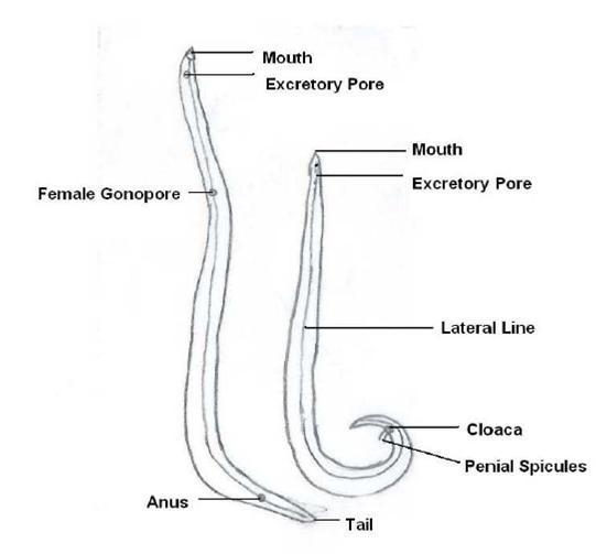
The worm is sexually diamorphic.
- Adult male: 15-30 cm in length, 3-4 mm in diameter, tail curved
- Adult female; 20-40 cm length, 2-6mm diameter, tail straight
Egg:
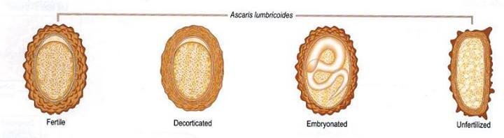
Ascaris egg is round or oval, 60*40 µm size, thick brown shell and have rough surface. It is the infective form of parasite.
- i) Un fertilized egg; large, more elongated (38-55*78-105) µm
- ii) fertilized egg; ovoid (35-50*50-70)µm, golden brown color
Life cycle:
The life cycle of Ascaris completes in single host. Human.
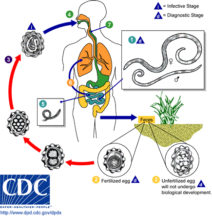
- Adult worm lives in small intestine
- Stages in life cycle:
Stage I: Eggs in faeces
- Sexually mature female produces as many as 200,000 eggs per day, which are shed along with faeces in unembryonated form. They are non infective.
Stage II: Development in soil
- Embryonation occurs in soil as optimum temperature of 20-25C with sufficient moisture and O2
- Infective larva develops within egg in about 3-6 weeks.
Stage III: Human infection and liberation of larvae
- Human get infection with ingestion of embryonated egg contaminated food and water
- Within embryonated state inside egg, first stage larvae develops into second stage larvae. This second stage larvae is known as Rhabtitiform larvae
- Second stage larve is stimulated to hatch out by the presence of alkaline pH in small intestine and solubilization of its outer layer by bile.
Stage IV: migration of larvae through lungs
- Hatched out larvae penetrates the intestinal wall and carried to liver through portal circulation
- It then travels via blood to heart and to lungs by pulmonary circulation within 4-7 days of infection.
- The larvae in lungs molds twice, enlarge and breaks into alveoli.
Stage V: Re-entry to stomach and small intestine
- From alveoli, the Larvae then pass up through bronchi and into trachea and then swallowed.
- The larvae passes down the oesophagus to the stomach and reached into small intestine once again.
- Small intestine is the normal habitat of Ascaris and it colonises here.
- Within intestine parasite molds twice and mature into adult worm.
- Sexual maturation occurs with 6-10 weeks and the mature female discharges its eggs in intestinal lumen and excreted along with faeces, continuing the life cycle.
- The life span of parasite is 12-18 months
Pathogenesis:
1. Mode of transmission:
- faeco-oral route, by contaminated vegetables or water.
2. Pathogenesis:
Infection of A. lumbricoides in man is known as Ascariasis. There are two phase in ascariasis.
Phase I: migrating larvae
- The migrating larvae causes pathological lesions. The severity of lesions depends upon the sensitivity of host, nutritional status of host and number of migrating larvae.
- During migration and molding through lungs, larvae may causes pneumonia with low grade fever, cough and other allergic symptoms.
Phase II: Adult worm
- Few worm in intestine produce no major symptoms and but some time give abdominal pain especially in children.
- The adult worm produce trauma in host tissue and the wandering adults may block the appendical lumen or common bile duct and even small intestine.
- Large number of adult worms affects the nutritional status of host by robbing the nutrition leading to malnutrition and growth retardation in children.
- The metabolites of living or dead worm are toxic and immunogenic.
- lumbricoides also produces various allergic toxin, which manifests fever, conjunctivitis and irritation.
Clinical manifestation:
Most of the Ascaris infection is asymptomatic.
- Symptomatic ascariasis; two types
- Intestinal Ascariasis
- Pulmonary Ascariasis
1. Intestinal ascariasis;
- Nausea
- Vomiting
- Colicky abdominal pain
- Abdominal distention
- Weight loss and diarrhea
- Malbasorption of nutrition
- Growth retardation
- Heavy worm in children leads to intussusception and total obstruction
- Complications: Appendicitis, Biliary colic and perforation of bile duct, Hepatomegaly
2. Pulmonary ascariasis;
- Transient eosinophilic pneumonitis (loeffler’s disease); elevated IgE
- Bronchospasm
- Dyspnea and wheezing
- Fever
- Non-productive cough and chest pain
Lab diagnosis:
- Specimen: stool, sputum
- Microscopy: examination of stool by saline emulsion or concentration by floatation methods employed to unembryonated egg
- X-ray
- Serodiagnosis: Indirect haemagglutination test, Immuno-fluorescence assay
- Ultrasonography and CT scan
- Other test: blood count shown peripheral eosinophilia
Treatment and prophylaxis:
- Mebendazole: drug of choice, (100mg twice a day for 3 days)
- Albendazole: 500mg single dose
- Pyrantel pamoate: single dose of 1omg/kg weight
- Piperazine citrate