
Trichomonas vaginalis: morphology, life cycle, pathogenesis, clinical manifestation, lab diagnosis, treatment, prevention and control
- Trichomonas vaginalis causes trichomoniasis. The infection is transmitted sexually.
- T. vaginalis trophozoites inhabitat the vagina in female, the prostate and seminal vesicles in male and urethra in both sexes.
Morphology of Trichomonas vaginalis
- It exists only in trophozoite stage, which is the infective form of parasite. Cystic stage is absent.
Trophozoite of T. vaginalis
- Shape and size:
- Trophozoite is pear-shaped and measures 7 mm to 23 mm in length.
- Motility:
- It shows twitching type of motility due to the presence of 5 numbers of flagella.
- The trophozoite is identified by its characteristic twitching motility.
- Flagella:
- There are four anterior free flagella, arising from a shallow depression in the anterior end of the body called periflagellar canal.
- The fifth flagellum curves back along the margin of the undulating membrane and is called the recurrent flagellum. It is not enclosed by the undulating membrane but lies in shallow groove in the free margin of the membrane.
- Costa:
- Just beneath the membrane lies the costa, which is unique to the trichomonads.
- The costa is a rigid cord, filamentous in appearance and is believed to support the undulating membrane.
- Axostyle:
- The parasite has an axostyle, which is a byaline rod like structure that runs through the entire length of the body and comes out at the posterior end.
- It is a part of the endoskeleton.
- Cytoplasm:
- The cytoplasm contains a large numbers of siderophilic granules and sometimes viral particles.
- Hydrogenosome:
- T. vaginalis is an anaerobic organism. Hydrogenosome is a modified mitochondira which produces energy by fermentation of sugars. In hydrogenosome enzyme of oxidative phosphorylation has been replaced by enzyme carrying out anaerobic fermentation.
Life cycle of Trichomonas vaginilis:
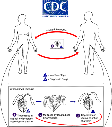
- Life cycle of T. vaginalis is simple. It is completed in a single host either male or female.
- The infection is transmitted sexually from a woman acting as a reservoir of infection to man.
- In the female, the parasite gets nourishment from the mucosal surface of the vagina and from the ingested bacteria and erythrocytes.
- T. vaginilis reproduces by longitudinal binary fission.
- It begins by division of the neuromotor apparatus and finally separation of cytoplasm into two daughter trophozoites.
- Trophozoites are the infective stages. On sexual contact, trophozoites are transmitted to male and localize in the urethra and prostate gland.
- These trophozoites probably undergo replication in the same way as seen in the vagina in females.
Trichomonas has following virulence factors:
- Protein liquids and proteases: They help in adherence of trophozoites to epithelial cells of the genitor-urinary tract.
- Lactic and acetic acid: it lower the pH of the vaginal fluid. The low pH of vaginal secretion is cytotoxic to epithelial cells
- Enzymes cysteine proteases: it is responsible for haemolytic activity of the parasite.
Mode of transmission;
- Venerally route by sexual contact with infected partners: It is the most common mode of transmission of infection in adolescents and adults.
- Occasionally non-venerally through fomites such as towels, toilet seats etc. and also from mud and water bath as well.
- By perinatal infection in some of female babies born to infected mothers during passage through the infected birth canal.
Pathogenesis and pathology of Trichomonas vaginalis
- T. vaginalis is an obligate parasite. It cannot survive outside the human body and needs close association of the vaginal, urethral or prostatic tissues for its survival.
- It is not an invasive parasite. It remains adherent to the mucosal epithelium of the vagina or urethra and causes super facial lesions. It infects squamous epithelium but not columnar epithelium.
- In pre-pubertal girls, the vaginal pH is more than 4.7, and the vaginal wall is thin and hypoestrogenic. As the girl attends puberty, the pH of the vagina lowers to less than 4.5. The vagina wall becomes thick and lactobacilli become the dominant flora of the vagina. The lactobacilli are important in protecting the vagina from infection by keeping the vaginal pH lower. The number polymorphonclear (PMN) leukocytes increase with an increase in the vaginal pH. These leukocytes are the key defense mechanisms of the host and they protect the host from the chomotactic substances produced by Trichomonas.
- Parasite causes degeneration and desquamation of the vaginal epithelium. Intra-cellular oedema and so called chicken-like epithelium is the most characteristic feature.
- Cellular atypia is a frequent finding. T. vaginalis destroys epithelial cells by direct cell contact and also by production of cytotoxic substance. The parasites combines with host plasma proteins, thus escaping from the lytic function of the alternative complement pathway and of the host proteineases.
Clinical manifestations of trichomoniasis
- T. vaginalis causes trichomoniasis ( or trichomonosis) which is the most common sexually transmitted disease in humans.
- Trichomonas produces a different spectrum of clinical manifestation in women and men. The disease is more often asymptomatic in men compared to women (unlike gonococcal infection where female mostly are asymptomatic)
- 1. Women :
- In asymptomatic women:
- Trichonomas causes vulvovaginitis and urethritis.
- The condition is asymptomatic in more than half of the cases.
- Symptomatic women:
- The incubation period varies between 5-10 days.
- Acute infection:
- Purulent abnormal vaginal discharge: Purulent abnormal vaginal discharge is the typical presentation of the trichomoniasis
- Strawberry cervix: mucosa lining of the vagina and ectocervix shows punchuate haemorrhage. This is called colpitis macularis which is very characteristics of trichomoniasis
- Vaginal erythema: vaginal epithelium are found fiery red and inflamed.
- Vaginal irritation
- Vulvovaginal soreness or irritation
- Dyspareunia
- Disagreeable odour and dysuria.
- Lower abdominal pain
- This acute stage may last for a week or month and often varies in intensity.
- * Note: The presence of T. vaginalis in pre-pubertal children may indicate sexual abuse of the children.
- 2. Men
- In men the infection is more often asymptomatic than symptomatic.
- Incubation period is usually 10 days but it can vary between 4-28 days.
- In symptomatic cases:
- T. vaginalis cause non gonococcal urethritis and rarely epididymitis
- Prostatis
- Superficial penile ulcerations at the pepuce.
- T. vaginalis is one of the important cases of non-gonococcal urethrits. It s characterized by pain in the urethra, testicular pain or a purulent to mucoid discharge.
- Most infection are intermittent and self-limiting. This possibly is due to trichomonicidal action of the prostatic fluid or flushing out of the flagellate mechanically from urethra during micturition.
3. Complications:
- In women, pelvic inflammatory disease is the most important complications.
- In Pregnant women infected with T.vaginalis are more likely to have premature rupture of membrane, premature birth and a pre-term or low birth weight baby.
- Prostatitis, epididymitis, urethral stricture and infertility are the common complications in men infected with T. vaginalis.
- T. vaginalis infection both in women and men is increasingly associated with the gonorrhea, Chlamydia and HIV infections.
Laboratory diagnosis of Trichomonas vaginalis
1. In women
- Specimens:
- vaginal discharge
- endocervical specimens
- *Endocervical specimens are not used for wet mount examination, because small numbers of parasites are present in this specimens. However the specimens can be collected for culture.
2. In men:
- Specimens:
These includes
- Urethral discharge
- Prostatic fluid
- Early morning first voided urine sediment
- Urethral swab before voiding urine in the morning
- Less commonly semen
Methods of examinations:
1. Microscopy
- Microscopy
of vaginal secretion shows-
- T. vaginalis trophozoites, which are identified by their jerky and twitching motility,
- large numbers of polymorphonuclear (PMN) cells and squamous epithelial cells.
- Bacteria flora consisting of rods or coccobacilli.
- Wet
mount examination;
- Trichomonas is the vaginal or ureteral discharge can be demonstrated by wet mount examination of saline.
- Wet mount is an easy, useful and economical method of parasitic diagnosis.
- *Iodine wet amount is not done because cystic stage is absent.
- Acridine-orange
staining:
- The smear made from vaginal or urethral discharge can be stained by acridine-orange.
- Acridine-orange staining is a rapid and accurate method to demonstrate trichomonads. It is sensitive as wet mount examination.
- Papanicolaou
stain:
- Vaginal smears can be stained by papanicolaou technique to detect trichomonads. Sensitivity varies from 60% to 70 % and is similar to that of wet amount.
- Direct
fluorescent antibody (DFA) staining:
- Direct fluorescent antibody staining of vaginal smear is more sensitive that the wet amount.
- This is rapid method for the diagnosis. Culture
2. Culture:
- Culture is considered as a gold standard for the diagnosis of T.vaginalis infection.
- It is the most sensitive method.
- In this method the specimen is inoculated in a medium and is cultured aerobically. In a positive culture, the actively motile trophozoites are demonstrated after 48 hours of incubation at 37°C.
3. Antigen detection:
- Enzyme-linked immunosorbent assay (ELISA)
- ELISA using a monoclonal antibody specific for a 65-k Da surface polypeptide of T. vaginalis is a very specific and sensitive method for detection of the parasite in vaginal secretion.
4. Serodiagnosis:
- It is non-specific test and has low sensitivity.
- These are of limited value in the diagnosis of trichomoniasis in individual cases.
5. Moleculr diagnosis
- DNA probes: the DNA probes have been used for identification of T. vaginalis DNA in vaginal and urethral secretions.
6. Other test
- Determination of vaginal pH:
- Vaginal pH is usually above 4.5 in trichomoniasis or in bacterial vaginosis but not in candidiasis.
- The vaginal pH is measured by Nitrazine paper method.
- Whiff test or Amine odor test:
- This test is positive in trichomoniasis and also in bacterial vaginosis.
Treatment of trichomoniasis
- Metronidazole is the drug of choice.
- It is given orally either in a single 2g dose or in a dose of 250mg three times daily for 7 days.
- This single dose therapy for trichomoniasis has the advantages of lower total dose, and good patient compliance. The drug produces a cure rate of 95%.
- Treatment of both sexual partners is recommended to prevent recurrence of infection and reduce the reservoir of infection.
In patients not responding to treatment with standard regimen, the following measures may be followed:
- The dosage may be increased.
- Metronidazole may be administered both in the oral and vaginal route or
- Materonidazole may be administered perenterally.
- T. vaginalis strains resistant to metronidazole have been isolated from some patients. In such resistant cases treatment with tinidazole has been found to be useful.
- Metronidazole is contraindicated in pregnancy due to its mutagenicity. In this situation, topical therapy with clotrimazole by applying vaginal suppositories in a dose of 100 mg, daily for 6 days is effective.
Prevention and control of trichomoniasis
- Detection and treatment of cases either male or female.
- Avoidance of sexual contact with infected partners
- Use of condoms
Trichomonas vaginalis: morphology, life cycle, pathogenesis, clinical manifestation, lab diagnosis, treatment, prevention and control