
Respiration, types of respiration and anatomy of Human respiratory system
Respiration
- Respiration is defined as the biochemical process by which the digested foods are oxidized liberating the energy. In the process, oxygen is utilized and carbon-dioxide is released.
- Overall respiration process involves three process.
- I. External respiration: it is a simple process of exchange of gases (O2 and CO2) between the respiratory surface and the environment.
- II. Transport of gases between the respiratory surface and the body tissue.
- III. Internal or cellular respiration: cellular respiration occur in mitrochondria of every cell, where digested food is oxidized releasing energy in the storable form ie. ATP. And when the cell require energy for vital activities, ATP broke down into ADP+ iP + energy.
Types of respiration
There are two types of respiration depending upon availability of oxygen
1. Aerobic respiration:
- It occur in the presence of Oxygen.
- The food is oxidized in the presence of oxygen in cellular level liberating CO2 and water along with energy in the form of ATP.
- Complete oxidation of 1 molecule of glucose liberate 38 molecule of ATP during glycolysis.
2. Anaerobic respiration:
- In the absence of oxygen, food is oxidized anaerobically (without utilizing o2).
- Anaerobic respiration is also known as fermentation as organic compounds are also produced as byproducts.
- One glucose molecule on anaerobic oxidation releases 2 ATP molecule only.
- Anaerobic respiration occur in deep seated tissue, in germinating seeds, parasites and bacteria.
Anatomy of Human respiratory system
The organs of the respiratory system are:
- nose (external nares and nasal chamber)
- Internal nares and pharynx
- larynx
- trachea
- two bronchi (one bronchus to each lung)
- bronchioles and smaller air passages
- two lungs and their coverings, the pleura
- muscles of breathing – the intercostal muscles and the diaphragm.
1. Nose (external nare and nasal chamber)
- Structurally the nose can be divided into the external portion which is in fact termed as the nose and the internal portions being the nasal cavities.
- The nose is the only visible part of the respiratory system, protruding from the face, and lying in between the forehead and the upper lip.
- Opening is known as nostril. The two nostrils are separated by nasal septum. The two nostril openings leading to two nasal chambers or cavities.
- The nasal cavity is the main route of air entry, and consists of a large irregular cavity divided into two equal passages by a septum.
- The posterior bony part of the septum is formed by the perpendicular plate of the ethmoid bone and the vomer. Anteriorly, it consists of hyaline cartilage.
- The nasal cavity is lined with very vascular ciliated columnar epithelium which contains mucus-secreting goblet cells.
The functions of nasal passage are:
- Prevent entry of dust particle into lungs
- Warm the incoming air entering the nasal cavity
- Moisten the dry air
- Olfactory receptor present in the roof of nasal cavity detect the smell
- Hold and sweep the microorgnisms entering the nasal chamber
2. Internal nares and pharynx
- The internal nares are the openings from the nasal cavity into the pharynx.
- The pharynx (throat) is a passageway that extends from the posterior nares, and runs behind the mouth and the larynx to the level of the 6th thoracic vertebra, where it becomes the oesophagus.
- Structurally the pharynx can be divided into three anatomical parts ie. nasopharynx (posterior to the nasal chambers), the oropharynx (posterior to the mouth), and the laryngopharynx (posterior to the pharynx).
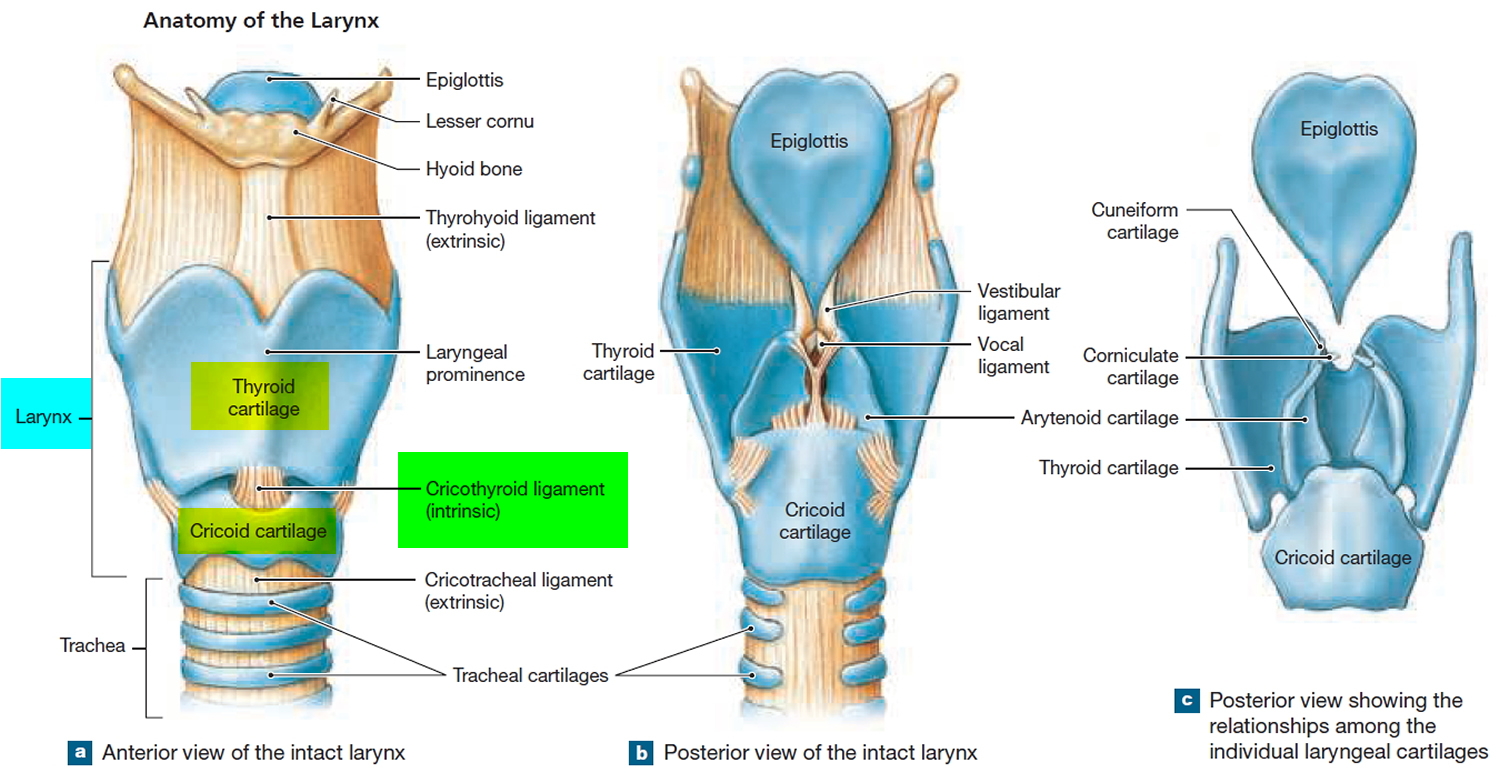
3. Larynx
- The larynx or ‘voice box’ links the laryngopharynx and the trachea. It lies in front of the laryngopharynx and the 3rd, 4th, 5th and 6th cervical vertebrae.
- Until puberty there is little difference in the size of the larynx between the sexes. Thereafter, it grows larger in the male, which explains the prominence of the ‘Adam’s apple’ and the generally deeper voice.
- The larynx is composed of several irregularly shaped cartilages attached to each other by ligaments and membranes.
- The main cartilages are:1 thyroid cartilage, 1 cricoid cartilage, 2 arytenoid cartilages and 1 epiglottis
- The vocal cords are two pale folds of mucous membrane with cord-like free edges, stretched across the laryngeal opening. They extend from the inner wall of the thyroid prominence anteriorly to the arytenoid cartilages posteriorly.
Sound production:
- When the muscles controlling the vocal cords are relaxed, the vocal cords open and the passageway for air coming up through the larynx is clear; the vocal cords are said to be abducted.
- Vibrating the vocal cords in this position produces low-pitched sounds.
- When the muscles controlling the vocal cords contract, the vocal cords are stretched out tightly across the larynx, and are said to be adducted (closed).
- When the vocal cords are stretched to this extent, and are vibrated by air passing through from the lungs, the sound produced is high pitched.
- The pitch of the voice is therefore determined by the tension applied to the vocal cords by the appropriate sets of muscles.
- When not in use, the vocal cords are adducted. The space between the vocal cords is called the glottis.
4. Trachea
- The trachea or windpipe is a continuation of the larynx and extends downwards to about the level of the 5th thoracic vertebra where it divides at the carina into the right and left primary bronchi, one bronchus going to each lung.
- It is approximately 10–11 cm long and lies mainly in the median plane in front of the oesophagus.
- The tracheal wall is composed of three layers of tissue, and is held open by between 16 and 20 incomplete (C-shaped) rings of hyaline cartilage lying one above the other.
- The rings are incomplete posteriorly where the trachea lies against the oesophagus.
- The tracheal mucosa consists of pseudo stratified, ciliated columnar epithelium, while its submucosa contains cartilage, smooth muscle, and seromucous glands.
5. Bronchus
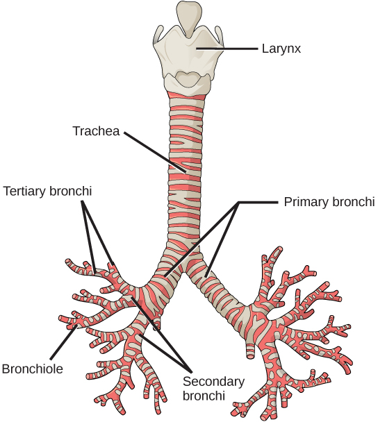
- The trachea divides into the two main bronchi (primary bronchi) ie. the right bronchus wider, shorter and more vertical than the left bronchus.
- The right bronchus: This is wider, shorter and more vertical than the left bronchus and is therefore more likely to become obstructed by an inhaled foreign body. It is approximately 2.5 cm long. After entering the right lung at the hilum it divides into three branches, one to each lobe. Each branch then subdivides into numerous smaller branches.
- The left bronchus. This is about 5 cm long and is narrower than the right. After entering the lung at the hilum it divides into two branches, one to each lobe. Each branch then subdivides into progressively smaller airways within the lung substance.
- The bronchial walls contain the same three layers of tissue as the trachea, and are lined with ciliated columnar epithelium. The bronchi progressively subdivide into bronchioles, terminal bronchioles, respiratory bronchioles, alveolar ducts and finally, alveoli.
6. Lungs
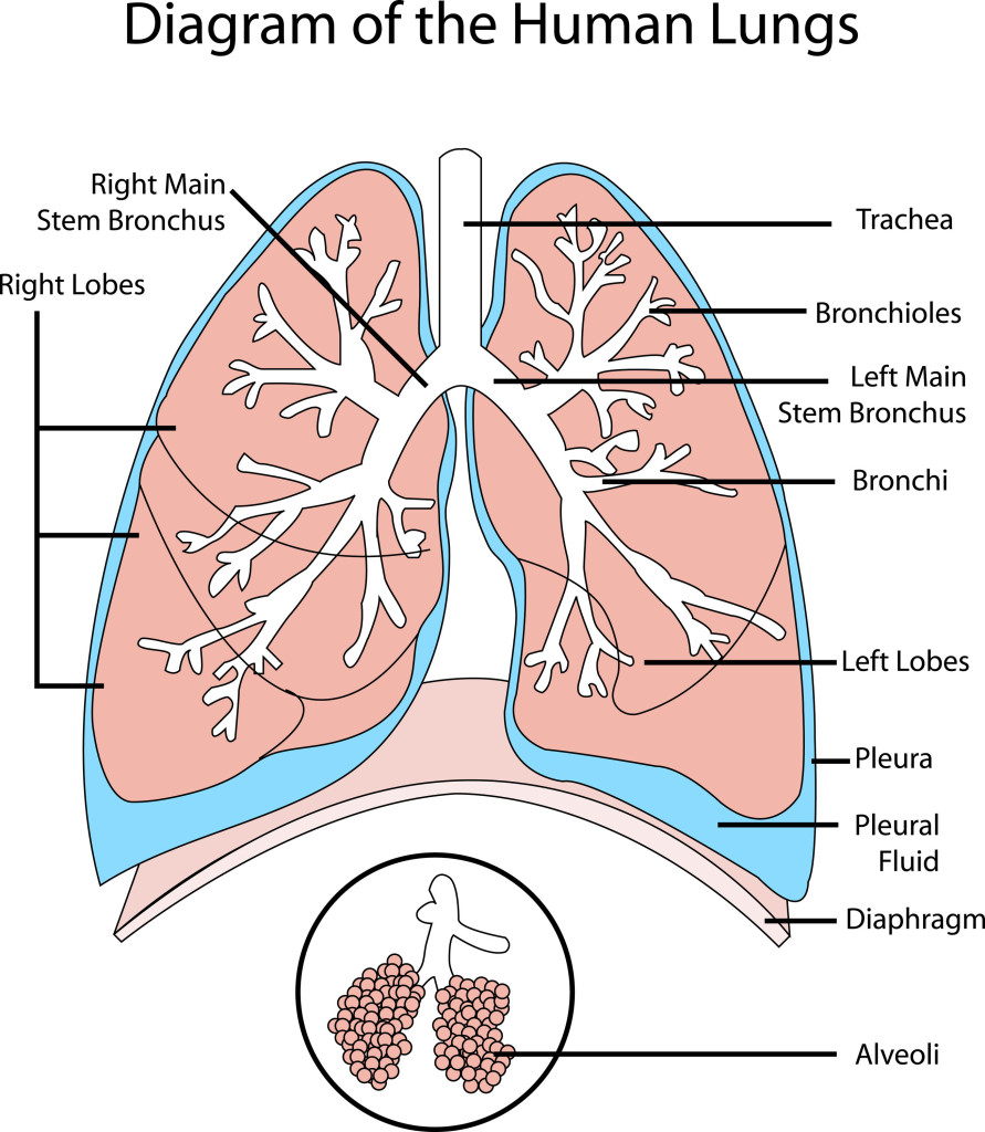
- There are two lungs, one lying on each side of the midline in the thoracic cavity. They are cone-shaped and have an apex, a base, a tip, costal surface and medial surface.
- The apex: This is rounded and rises into the root of the neck, about 25 mm above the level of the middle third of the clavicle. It lies close to the first rib and the blood vessels and nerves in the root of the neck.
- The base: This is concave and semilunar in shape, and lies on the upper (thoracic) surface of the diaphragm.
- The costal surface: This is the broad outer surface of the lung that lies directly against the costal cartilages, the ribs and the intercostal muscle.
- The right lung is divided into three distinct lobes: superior, middle and inferior. The left lung is smaller because the heart occupies space left of the midline. It is divided into only two lobes: superior and inferior. The divisions between the lobes are called fissures.
- Lungs are enclosed in the pleural cavity lined by transparent pleural membrane. The pleura consists of a closed sac of serous membrane which contains a small amount of serous fluid.
- The lung is pushed into this sac so that it forms two layers: one adheres to the lung called visceral pleura and the other to the wall of the thoracic cavity called parietal pleura.
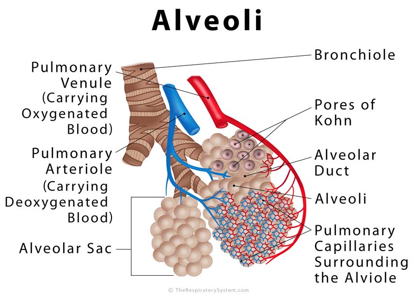
7. Alveoli
- Within each lobe, the lung tissue is further divided by fine sheets of connective tissue into lobules.
- Each lobule is supplied with air by a terminal bronchiole, which further subdivides into respiratory bronchioles, alveolar ducts and large numbers of alveoli (air sacs).
- There are about 150 million alveoli in the adult lung. It is in these structures that the process of gas exchange occurs.
- As airways progressively divide and become smaller and smaller, their walls gradually become thinner until muscle and connective tissue disappear, leaving a single layer of simple squamous epithelial cells in the alveolar ducts and alveoli.
- These distal respiratory passages are supported by a loose network of elastic connective tissue in which macrophages, fibroblasts, nerves and blood and lymph vessels are embedded.
- The alveoli are surrounded by a dense network of capillaries.
- Exchange of gases in the lung (external respiration) takes place across a membrane made up of the alveolar wall and the capillary wall fused firmly together. This is called the respiratory membrane.