
Onchocerca volvulus
- Onchocerca volvulus derived its name from two Greek word, onkos-hook, cercos-tail, hence meaning ‘hooked tailed’.
- It is a nematode that causes onchocerciasis or ‘River blindness’, mostly in Africa.
- It is one of the leading cause of blindness in many parts of the world, hence it is popularly known in America as ‘blinding filaria’. Besides this it also causes cutaneous filariasis.
- O. volvulus along with other filarial nematode share an endosymbiotic relationship with bacteria Wolbachia. In the absence of Wolbachia, larva development of O. volvulus is disruptred.
Habitat
- Adult worms are found in the nodules in the subcutaneous tissues, when they become encapsulated by the host immune reactions.
- Microfilaria are found in the nodules and the dermal layers of the skin and also in the blood and eye during heavy infection.
Morphology
i. Adult worm:
- Adult worm are white, opalescent and transparent.
- Microscopically the cuticle is seen and it is more prominent in female than in male.
- The posterior end or tail is curved hence the name Onchocerca.
- The adult worms of either or different sexes are usually coiled together in the subcutaneous nodules.
- The intestinal cells are filled with concentric spherules and the intestinal lumen is reduced.
- The worms usually live 10 or more years in their host.
- Male:
- Male measures 19-42cm in length and 0.13-0.21 mm in diameter and is curved and bulbous.
- The posterior end of male is coiled ventrally which bears 4 pairs of ad-anal and 6 or 8 pairs of post-anal papillae.
- Male has also two spicules of unequal length
- The cuticle of male consists more layers and its epicuticle is wrinkled
- Female:
- Female measures 33-50 cm in length and 0.27-0.4 mm in diameter.
- The vulva is located behind the posterior extremity of the oesophagus.
- The cuticle of female consists of fewer layers and the epicuticle forms protuberances.
ii. Microfilariae
- They are found in the skin.
- They are unsheathed and non-periodic.
- They measure 300 µm in length and 6-8 µm in breadth.
- Anterior end is slightly enlarged and tail is pointed. But both the anterior and posterior ends are nucleus free.
iii. Infective larva
- It is found in the mouth parts of the black fly (vector; Simulium spp)
- They measures 10 mm in length.
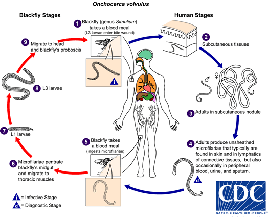
Life cycle of Onchocera volvulus:
- Life cycle completes in two host.
- Definitive host: Human
- Intermediate host: Black fly of genus Simuliaum
- Human acquires infection by the injection of infective larva into the skin by the bite of female black fly.
- The larvae enter the skin through the punctured wound and migrate to the subcutaneous tissue in which they moult and develop into adult male and female worms.
- As the infection progresses these worms induce the formation of fibrous nodule, in which they remain encapsulated.
- After a pre-patent period of 7-34 month, the female worms after being fertilized by males produces unsheathed microfilariae.
- The microfilariae then migrate out of the fibrous nodule to enter the dermis of the skin and the connective tissue. They also migrate to the eye and other organs of the body but rarely to the circulation.
- During blood mean from infected person, microfilaria enter the black.
- In the stomach of black fly, these microfilaria penetrate the gut wall and migrate through the hemocoel to the thoracic muscles and moult twice over a week.
- Then they migrate to the black fly’s proboscis and develop into infective larva which affect another human when they take the blood meal and the cycle is repeated.
Mode of transmission:
- Infected man is the only source and reservoir of infection.
- Man to man infection is acquired by the bite of infected black flies of genus Simulium.
Pathogenesis of Onchocerca volvulus:
- Adult worms in the subcutaneous tissue produce varying degree of inflammation and nodules.
- These subcutaneous nodules usually appear 3-4 month after the infection. Subsequently, adult worms may be surrounded by a granulomatous inflammatory reaction followed by formation of granuloma fibrosis or calcification.
- The microfilaria are believed to cause many of the disease manifestation of onchocerciasis.
- Trauma caused by living microfilaria and hypersensitivity reactions of the host to dead microfilaria is suggested to be responsible for pathological lesions in the eye and also an inflammatory dermatitis in the skin.
Clinical symptoms of Onchocerca infection:
- Onchocerca volvulus causes onchocerciasis in human.
- The pathological lesions in the skin and eye result from a hyper sensitivity reaction to the dead or dying microfilaria.
- The incubation period in man is about 1 year.
1. Lesions:
- The pathological lesions can be divided into two groups.
i. Skin lesions
- Skin involvement typically consists of intense itching, swelling and inflammation.
- Acute parasitic rashes
- Lichenified onchodermatitis spotted depigmentation of the skin leads to “Leopard skin appearance, usually an anterior lower leg.
- Skin atrophy and loss of elasticity resulting in “Lizard at skin “ appearance
ii. Onchocercoma:
- Onchorcercoma or subcutaneous nodules are the typical pathological lesions.
- These nodules are visible or palpable and contains the adult worm.
- Subsequently the adult worms may be surrounded by granulomatous inflammatory reaction followed by the formation of granuloma, fibrosis or calcification.
- The nodules usually appears 3-4 month after the infection.
- These lesions are firm, smooth and rubbery in consistency. These are freely mobile and attached to the underlying tissue. The size varies from a few mm to several cm.
Other infections:
i. Lymphadenopathy:
- The microfilaria can cause the inflammation of lymph nodes, especially in the inguinal and femoral areas.
- The enlarged lymph nodes along with loss of tissue elasticity can lead to a condition known as ‘Hanging groin”.
ii. Ocular lesions and blindness:
- It is particularly seen in persons with nodules on head or face.
- They result from the presence of microfilaria which may be found moving about in the substantia propria of the cornea and also in the anterior chamber.
- The clinical manifestation consists of simple conjunctivitis, small round opacities and pannus in the anterior guardant of the cornea. Later there may be iridocyclitis, secondary glaucoma and papilitis (optic atrophy, eventually leading to blindness.
iii. Complications
- Secondary bacterial infection of the skin lesions
- Blindness is a serious complications of ocular lesions
iv. Mazzoki reaction:
- It is a symptoms complex seen in patients after undergoing treatment of the onchocerciasis with Diethylcarbamazine (DEC).
- Mozzoki reaction generally occurs within 7 days of treatment.
- The reaction can be life threatening and are characterized by fever, urticaria, swollen lymph nodes, tachycardia, hypertension, arthralgias, oedema and abdominal pain.
Laboratory Diagnosis of Onchocerca volvulus:
- Diagnosis is based on demonstrating the microfilariae in the shaved piece of skin and the adult worms inside the excised nodules.
Specimen:
- Skin snips, excised nodules
Microscopic examination of skin snip:
- Small area of the skin about 3 mm in diameter is raised with the tip of needle and then cut off with the razor blade.
- The skin snips are then immersed in the physiological saline on a slide or in a well of a micro filtration plate.
- These are kept at room temperature for 30-60 min, during with microfilaria emerge out of the skin snips to the physiological saline.
- The preparation is examined microscopically for the microfilariae.
- This method is specific and most accurate. However it is less sensitive for detecting early or mild infections.
Sero-diagnosis:
- The serological test are useful for the diagnosis of cases in which microfilaria are not demonstrated in the skin snips.
- ELISA is the most sensitive method to detect antibodies against specific Onchocercal microfilarial antigens.
- A rapid card test
Molecular diagnosis:
- PCR
Other diagnosis test:
- USG: Ultrasound is useful to detect the deep onchocercomas and vitreous changes in the eye.
- DEC patch
test:
- It is also known as Mazzoki patch test.
- When the drug patch is placed on the skin, the infected persons show localized pruritus and urticaria as a reaction to dead microfilaria within 24 hours.
- Skin
scratches test
- This is a recent approach as a minimally invasive and painless alternative to use of skin snips.
- This methods involves careful removal of the superficial layer of the epidemics by carefully scraping the skin with the blunt edge of disposable lancet.
Treatment of Onchocerca volvulus:
- IVERMECTIN:
- It is a drug of choice for Onchocerca infection.
- Dose: single oral dose of 150 mg per kg weight
- HETRAZAN and SURAMIN
- Nodulectormy: Surgical removal of detectable nodules wherever possible.
Prevention and control
- Control of the vector by destruction of the larva of black flies by applying larvicides in their breeding places.
- Personal protection by wearing protective clothing.
Epidemiology and geographical distribution of Onchocerca volvulus:
- Onchocerca infection are found in tropical climate
- Onchocerciasis is endemic in 37 countries in Africa, Latin America and Middle East.
- Scattered foci of endemic areas occur in Mexico and Guatemala of Central America, Brazil, Venezuela and Yemen in middle East