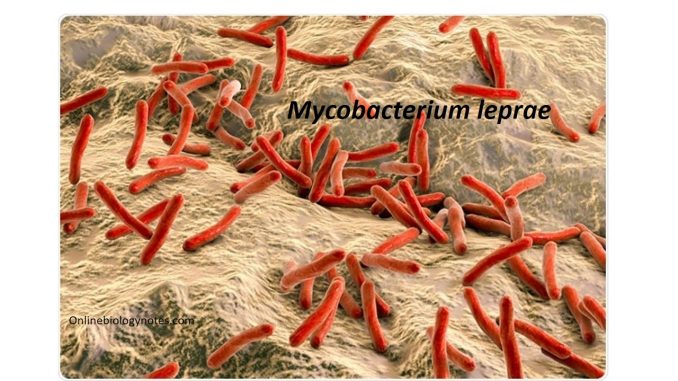
Mycobacterium leprae
- Mycobacterium leprae is the only pathogenic bacteria of man that has not yet been cultivated in vitro and thus fails to fulfill the Koch’s postulates.
- The lepra bacillus was first observed by Hansen in 1873 so it is also called Hansen’s bacilli.
General characteristics:
- M. leprae is a straight or slightly curved rod, (1-8 mm X 0.2 -0.5 mm) in size with parallel sides and rounded ends.
- They are arranged singly, in parallel bundles in a packet or in globular masses.
- Polar bodies and other intracellular elements may be present.
- The bacteria show considerable morphological variations. They exhibit cubical, lateral or branching forms.
- They are non-motile, non-spore forming.
- They are Gram positive and stains readily than M. tuberculosis.
- They are acid fast bacilli but less thus 5 % H2SO4 is used for decolonization during staining procedures.
- However, they are not alcohol fast.
- In stained smears, the live leprae bacilli appear solid and uniformly stained whereas the dead appear fragmented and granular.
- The bacilli are seen singly or in groups, intracellularly or lying free outside the cell.
- They frequently are bound together by a lipid like substances the glia.
- These masses of bacteria lying together is called ‘globi’ that present a ‘cigar bundle’ appearance.
- The globi appears in Virchow’s ‘lepra cell’ or ‘foamy cell’ which are large undifferentiated histocytes.
- M. leprae is an obligate intracellular parasite that multiplies preferentially in tissues at cooler temperature.
Habitat:
- M. leprae is found in large number in infected nasal secretions of patients with lepromatous leprosy.
Virulence factors:
The virulence factors exhibited by M. leprae includes:
- Fibronectin:
- Enhances the attachment and ingestion of M. leprae by epithelial and Schwann cell lines.
2. Secreted proteins:
- Two secreted proteins, early secretory Antigenic target (6 Kda) and culture filtrate protein 10 are thought to separately form heterodimers that prevent fusion of the phagosome and lysosome and thus enable mycobacteria to grow within immune cells.
3. Lipoarabinomannan (LAM):
- LAM is a lipoglycan in the cell wall.
- It is thought to hinder macrophage activity as well as provide resistance to oxidative radicals.
- It inhibits antigen presentation and prevents release of IFN-8, a macrophage activating cytokine.
4. Phenolic glycolipid -1 (PGL-1):
- PGL-1 is a prominent surface lipid found on the outermost layer of the cell wall.
- It binds to C3 component of the complement which leads to phagocytosis mediated by CR1, CR3 and CR4 receptors found on the cell surfaces.
- PGL-1 protects the lepra bacillus once inside the phagocytic cells, from oxidative killing by macrophages by removing hydroxyl radicals and superoxide anions.
5. Intracellular bacilli:
- The intracellular location of the bacteria makes them resistant to killing by phagocytes.
6. Mycolic acid:
- The lipid rich waxy cell wall containing mycolic acids also acts as an important virulence factor.
