
Human Eye: Anatomy, parts and structure
- The eye is the photo-receptor organ.
- Size and shape: Human eye is spherical about 2.5 cm in diameter.
- Location: it is situated on an orbit of skull and is supplied by optic nerve.
- There are 6 sets of muscles attached to outer surface of eye ball which helps to rotate it in different direction.
- Four sets of these muscles are straight muscles; superior, inferior, medial and lateral rectal muscle and two sets are oblique muscles; superior and inferior oblique muscles.
- Structurally two eyes are separated but some of their activities are coordinated so that they functions as a pair.
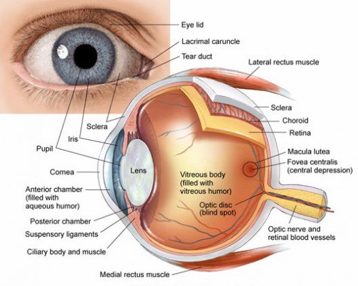
Anatomical structure of Eye
Eye ball consists of three layers
- Outer fibrous layer: Sclera, cornea and conjunctiva
- Middle vascular layer: ciliary body, choroid and iris
- Inner layer: retina
I. Outer fibrous layer:
It consists of following parts.
1. Sclera:
- It is outermost supporting layer consists of thick membrane of tough fibrous connective tissue.
- It covers 5/6 parts of eye ball.
- It maintains the shape of eye and provide attachment to the extrinsic muscle of eye
2. Cornea:
- It is a thin transparent front part of sclera.
- It forms a slight bulge at the front and covers an anterior 1/6 part of sclera.
- Cornea is avascular and absorbs oxygen from air.
- It refracts light to focus on retina.
3. Conjunctiva:
- It is a thin transparent layer that covers the cornea.
- It is formed of single layer of stratified squamous epithelium
- It protects the cornea.
II. Middle vascular layer:
It consists of following parts:
1. Choroid:
- It is thick vascular and pigmented layer situated below sclera.
- The pigmented cells absorbs light and prevent it from being reflected.
- The function of choroid is to provide nutrition and to prevent reflection of light.
2. Ciliary body:
- These are attached to choroid and present at the junction of sclera and cornea.
- It consists of two sets of ciliary muscle and suspensory ligament.
- Ciliary body is attached to lens and holds it in position
- Its function is to change the shape of lens by contraction or relaxation of muscle
3. Iris:
- It is muscular, pigmented and opaque diaphragm which hangs in the eye ball in front of lens.
- It has small circular opening called pupil.
- It has two types of muscles; circular and radial muscle. The movement of these muscles control the size of pupil.
- Pigment in iris gives color to eye.
- Iris control the amount of light entering into eye by controlling the size of pupil.
III. Inner layer:
It consists of photoreceptor cells and photo sensitive elements.
1. Retina:
- Retina is innermost layer.
- Neuroretina contains highly specialized photoreceptor nerve cells; rods and cones
- Each eye ball has 125 millions of rod cells and 7 millions of cone cells.
- Small depression in retinal wall is called Fovea centralis which contains only cone cells.
- Fovea centralis is highly sensitive to light and forms magnified image and give sharp and acute vision.
- The optic nerve enter retina at a point called blind spot. It does not contains any rods or cone cells. It is least sensitive to light and forms no image when light falls on blind spot
Rod cell:
- rods are sensors for perception of black to white shades
- Night vision is almost rod vision.
- It function in dim light
- Contains a photosensitive pigment rhodopsin formed from vitamin A.
Cone cell:
- Cones are sensors for perception of colors.
- It functions in bright light and differentiate colors.
- Contains a photosensitive pigment iodopsin.
Eye lens and chambers
1. Eye Lens:
- It is a large, flexible, transparent biconvex and fibrous crystalline body situated behind iris.
- Lens is enclosed in a transparent elastic capsule.
- Ciliary muscles control the thickness of lens and its power of accommodation.
- It forms the image of the object on retina.
- Lens separates the eye ball into two chamber
i. Aqueous chamber
ii. Vitreous chamber
Aqueous chamber:
- It is a smaller fluid filled chamber between cornea and lens.
- It is filled with aqueous humour containing aminoacids, glucose, ascorbic acid, hyaluronic acid and respiratory gases.
- The aqueous humour nourishes the lens and cornea and refracts light rays to focus on retina.
Vitreous chamber:
- It is a larger fluid filled chamber between lens and retina.
- It is filled with gelatinous vitreous humour containing salts and muco proteins
- It supports retina and refracts light to focus on retina.