
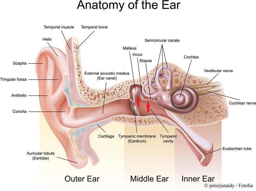
Human Ear: Structure and Anatomy
- The anatomy of ear consists of external ear, middle ear and inner ear.
- External ear (outer ear): Auricle (pinna) and external auditory meatus
- Middle ear: tympanic membrane and auditory ossicles
- Inner ear: vestibules, semicircular canal, cochlea
External ear:
External ear is composed of auricle and external auditory canal (meatus).
1. Auricle (pinna)
- Auricle is composed of thin plate of elastic cartilage covered by layer of skin.
- The funnel like curves of auricle collects sound wave and direct them to middle ear.
- The deepest depression called concha which is partly covered by two small projection; tragus in front and antitragus behind.
2. External auditory meatus
- External auditory meatus is slightly curved canal of about 2.5 cm ling extending from floor of concha to tympanic membrane (ear drum).
- The meatus is lined with skin continuous with auricle. It contains two glands; sebaceous gland and ceruminous gland.
- Ceruminous gland are modified sweat gland that secretes cerumen (wax).
3. Tympanic membrane:
- It is oval bluish grey membranous structure located on medial part of auditory meatus.
- It separates external and middle ear.
- It is a stretchable organ capable of vibrating.
- It receive sound wave and amplify into appropriate magnitude.
Middle ear:
Middle ear is small chamber between tympanic membrane and inner ear. It consists of tympanic cavity and contains ear ossicles.
1. Tympanic cavity:
- Tympanic cavity is a narrow irregular air filled space in temporal bone. It is separated from external ear by tympanic membrane and medially from inner ear by bony wall.
- It has two opening; oval window and round window.
- In the anterior wall of tympanic cavity is an auditory tube, commonly called as Eustachian tube.
2. Eustachian tube
- Eustachian tube leads downward from tympanic cavity to nasopharynx.
- It is about 4 cm long.
- The mucus membrane lining the nasopharynx is also continuous with membrane of tympanic cavity through eustachiantube. As a result of which infection from nose or throat may spread to middle ear causing Otitis media.
- The main purpose of Eustachian tube is to maintain equal air pressure on both side of tympanic membrane by permitting air to pass from nasal cavity to middle ear.
3. Ear ossicles:
- The three ear ossicles (malleus, incus and stapes) form a chain of lever extending from tympanic membrane to inner ear.
- The ear ossicles transmit sound wave from ear drum to inner ear.
- Ear ossicles communicate the ear drum with internal ear through fenestra ovalis ( oval window).
- The ear ossicles are;
- Malleus: it is hammer shaped bone whose handle is in contact with tympanic membrane and the head form movable joint with incus
- Incus: it is middle anvil shaped bone.
- Stapes: it is medial stirrup shaped bone. It head articulate with incus and foot plate fits into oval window. Stapes is the smallest bone in human body.
Inner ear:
The inner ear is also called as labyrinth because of its intricate structure of interconnecting chamber and passage.
It consists of two main structural parts one inside the other.
- Bony labyrinth
- Membranous labyrinth
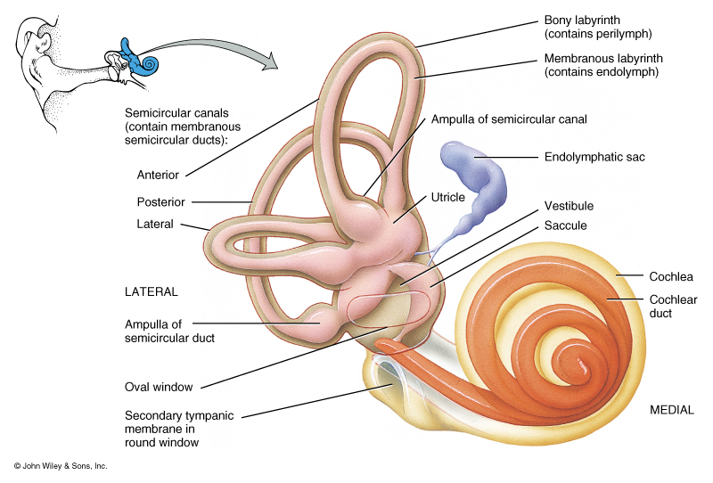
1. Bony labyrinth:
- It is a series of hollow channels. It is filled with perilymph.
- Bony labyrinth consists of vestibule, three semicircular canal and spirally coiled cochlea.
2. Membranous labyrinth:
- It is surrounded by bony labyrinth. It is filled with endolymph and also contains the sensory receptors for hearing and equilibrium.
- Membranous labyrinth consists of three semi-circular ducts as well as utricle, saccule and cochlear duct, all are filled with endolymph enclosed by bony labyrinth.
- It also contains sensory receptors (cristae, ampullaris maculae and organ of corti).
- Semi-circular dusts are located within semicircular canal of bony labyrinth.
- Perilymph is located in space between duct and bony wall of semicircular canal.
Vestibule:
- Vestibule is the expanded part nearest the middle ear.
- It has two sacs; larger upper utriculus and smaller lower sacculus.
- Utriculus and sacculus is connected by utriculosaccular duct.
- The sensory sport (macula) is present in both utriculus and sacculus.
- The macula consists of otolith membrane having otolith (small crystal of CaCO3) which concerned with balancing of body.
Semicircular canal:
- It is associated with equilibrium or balancing not for hearing.
- There are three semicircular canals arises from utriculus; anterior, posterior and lateral canals
- The anterior and posterior canals opens at one end to form common duct called crus commune. One end of each semicircular canal is swollen to form ampulla.
Cochlea:
- It is spiral shaped resembling snail’s shell, wounded 2 ¾ times.
- It is the main hearing organ.
- It is connected with cerebrum by vestibulo-cochlear nerve.
- Cochlea is divided into 3 spiral fluid filled chamber.
i. Scala vestibuli:
- It communicate with vestibule.
- It contains perilymph
ii. Scala tympani:
- It ends at round window of tympanic cavity.
- It contains perilymph.
iii. Scala media or cochlear duct:
- It lies between scala vestibuli and scala tympani.
- It contains endolymph.
- Scala media or cochlear duct is separated from scala vestibuli by vestibular membrane and from scala tympani by basilar membrane.
- The basilar membrane has organ of corti formed about 24000 receptor auditory cells.
Organ of corti:
- It is the organ for hearing which is rested on basilar membrane.
- The organ of corti present within scala media of cochlea that receive and conduct sound stimulus.
- Organ of corti is an organized structure consisting of hair cells and supporting cells.
- Hair cells are arranged in rows along the length. The outer hair cells are arranged in three rows and inner hair cells are arranged in single row.
- Each outer and inner hair cell have sensory hair which are specialized microvilli.