
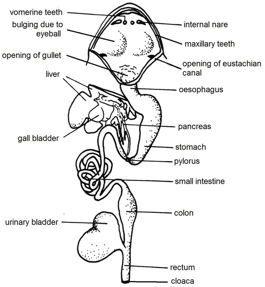
Digestive system of Frog: parts and functions
- Digestive system consists of digestive tract or alimentary canal along with the associated digestive glands.
- Alimentary canal:
- Alimentary canal of frog is complete.
- It is long and coiled tube. The tubes have varying diameter.
- It extends from mouth to cloaca.
- It consists of:
- Buccal cavity
- Pharynx
- Oesophagus
- Stomach
- Small intestine
- Large intestine
- Cloaca
1. Mouth:
- It is the beginning to the alimentary canal.
- Mouth is a very wide gap. It extends from one side of the snout to the other.
- Two bony jaws bound the mouth, and the jaws are covered by immovable lips.
- The upper jaw is fixed.
- The lower jaw is flexible i.e. it can move up and down to close or open the mouth.
2. Buccal cavity of frog:
- Mouth opens into buccal cavity.
- Buccal cavity is large, wide and shallow.
- It has ciliated columnar epithelial lining that contains mucous glands.
- These mucous glands secrete mucus that helps in lubricating the food.
- Frog lacks salivary glands.
- Teeth:
- The lower jaw lacks teeth.
- However, teeth occur in a row of either side on the premaxillae and maxillae bones of the upper jaw. The teeth are backwardly pointed.
- Vomers (two small bones in the roof of the mouth) also consists of two groups of vomerine teeth.
- The function of teeth is to simply hold the prey and prevent it from slipping out.
- Teeth are not meant for chewing.
- The nature of teeth is homodont (similar), acrodont (not set in a socket).
- But teeth are attached to the jaw bone by a broad base made of a bone-like substance.
- The crown is the free part of tooth.
- It is made up of dentine (a hard ivory-like substance), which is traversed by numerous fine canals or canaliculi.
- Enamel covers the tip of the crown.
- Enamel is a very hard, resistant and glistening substance.
- Tooth contains a central pulp cavity open at the side.
- It is filled with a soft nourishing pulp, containing connective tissues, blood vessels, nerve and odontoblast cells that produces new material for the growth of tooth.
- Frog are polyphyodont in nature, i.e. teeth is replaced several times in life.
- Tongue:
- In frogs, tongue is large, muscular, sticky and protrusible.
- It lies on the floor of mouth cavity.
- The anterior end of tongue is attached to the inner border of lower jaw.
- The posterior end is free and bifid.
- This free end can be flicked out and retracted immediately after catching the prey.
- The slimy surface of tongue facilitates in capturing the prey.
- The change of pressure in large sublingual lymph sac causes the protrusion of tongue.
- Internal nostrils:
- Just in front of vomerine teeth, the roof of buccal cavity contains anteriorly, a pair of small openings of internal nares.
- By these internal nares, the nasal cavities open into buccal cavity.
- These serves in respiration.
- Bulging of orbits:
- The roof of buccal cavity shows two large oval and somewhat pale areas, behind the vomerine teeth. These areas are the bulging of eye balls.
- In course of swallowing the food, frog depresses the eyes.
- This causes the orbits to bulge inwards which in response pushes the food towards the pharynx.
3. Pharynx:
- Posteriorly, the buccal cavity reaches short pharynx without any clear demarcation.
- So, sometimes these are termed as single bucco-pharyngeal cavity.
- Several apertures open into pharynx.
- A median elevation on the floor carries the glottis.
- Glottis is a longitudinal slit like aperture.
- The glottis leads to the laryngo-tracheal chamber.
- A wide eustachian aperture is present on either lateral side in the roof.
- This aperture opens into the middle ear.
- In male frogs, on the floor of pharynx, the small opening of a vocal sac is present on either side near the angle of two jaws.
- Now, the pharynx tapers behind to lead to esophagus through the gullet.
- Gullet is the wide opening that leads to Oesophagus.
4. Oesophagus:
- Oesophagus is a short, wide, muscular and highly distensible tube.
- Its mucous epithelial lining is folded longitudinally and contains some mucous glands.
- During the passage of food, its expansion is allowed by longitudinal foldings.
- An alkaline digestive juice is secreted by the glandular lining of oesophagus.
- Oesophagus enlarges to join with stomach in the peritoneal cavity.
5. Stomach:
- Stomach is present on the left side in the body cavity.
- It is attached to the dorsal bodywall by a mesentery termed as mesogaster.
- It is around 4 cm long, broad and slightly curved bag or tube with thick muscular walls.
- The anterior part is large, and broad. It is called as cardiac stomach.
- The posterior part is short and narrow. It is called the pyloric stomach.
- Several prominent longitudinal folds are present in the inner surface of the stomach.
- It allows the distension of stomach when food is received.
- Its mucous epithelium has multicellular gastric glands.
- These glands secrete the enzyme pepsinogen and unicellular oxyntic glands, secreting hydrochloric acids.
- The pyloric end of stomach is slightly constricted.
- Pyloric valve guards its opening into small intestine.
- Pyloric valve is a circular ring like sphincter muscle.
- Stomach serves for storage as well as digestion of food.
6. Small intestine:
- Small intestine is a long, coiled and narrow tube.
- It is about 30cm long, and is attached mid-dorsally to bodywall by mesenteries.
- It comprises of two parts:
- A small anterior duodenum
- A much longer posterior ileum
- Besides, intestinal glands, the mucosal lining of the small intestine consists of two types of cells.
- They are:
- Goblet cells:
- Large cells containing oval vacuoles and granular substances which produces mucus.
- Near the base of the cell, nucleus is present.
- Absorbing cells:
- Small cells with nuclei near the base.
- Duodenum:
- Duodenum runs ahead being parallel to stomach and forms a shape like U.
- It receives a common hepatopancreatic duct.
- Liver and pancreas bring bile and pancreatic juice respectively.
- Low transverse folds are formed by the internal mucous lining.
- Ileum:
- Ileum is the longest part of alimentary canal.
- Before enlarging posteriorly to join rectum, it makes several loops.
- The internal mucus lining forms many longitudinal folds.
- However, as in case of higher vertebrates, there are no true villi and definite glands and crypts.
- In the small intestine, digestion of food and absorption of digested food takes place.
7. Large intestine or rectum:
- Large intestine is short, wide tube about 4cm long.
- It runs straight behind to open into cloaca by anus.
- The opening is guarded by an anal sphincter.
- The inner lining of large intestine forms numerous low longitudinal folds.
- Itserves for the re-absorption of water and the preparation and storage of faeces.
8. Cloaca:
- It is the small terminal sac-like part.
- The anus and the urinogenital apertures open into cloaca.
- Cloaca opens to outside by the vent or cloacal aperture, lying at the hind end of body.
Digestive glands of frog:
- Keeping aside gastric glands and intestinal glands, two large glands that are linked with the alimentary canal of frog are the liver and the pancreas.
- Liver:
- The largest gland in the body of vertebrate is the liver.
- It is reddish-brown in colour.
- It is multi-lobed gland and lies close to the heart and lungs.
- 3 lobes are present in the liver of frog i.e. right, left and median.
- Liver consists of innumerable polygonal cells that secretes bile.
- Bile is a greenish alkaline fluid.
- Bile is stored in the thin-walled sac called as gall bladder.
- Gall bladder is large, spherical, and greenish in color.
- A common bile duct is formed when cystic ducts from gall bladder and hepatic ducts from liver lobes combines.
- It runs through pancreas and joins the pancreatic duct to form a hepatopancreatic duct.
- Now, it ultimately opens into duodenum.
- Bile lacks any digestive ferments and only emulsifies fats.
- Thus, liver is not a true digestive gland.
- Pancreas:
- Pancreas of frog is much branched, irregular flattened and is yellow in color.
- It lies in the mesentery between stomach and duodenum.
- It carries out both exocrine and endocrine function.
- The endocrine part is formed by scattered islets of Langerhans. It produces insulin hormone which is related to sugar metabolism.
- The exocrine part secretes pancreatic juice. This juice contains of several digestive enzymes.
- Since pancreas lacks independent duct, the pancreatic juice reaches the duodenum through the hepatopancreatic duct.
Physiology of digestion in frog:
- Being strictly carnivorous, frog feeds on insects, worms, crustaceans, molluscs, small fish and even small frogs and tadpoles.
- The prey is caught by rapid flicking of tongue and is swallowed as a whole.
- The food is now passed to stomach.
- As salivary glands are absent in case of frogs, the food is lubricated by the mucus secreted from the lining of bucco-pharyngeal cavity and oesophagus.
- The wave of contraction of the muscular wall of oesophagus pushes food down, it is called as peristalsis.
- Gastric digestion:
- Food remains in the stomach for upto 2-3hrs, which is sufficient time.
- Gastric juice is secreted by the gastric glands of stomach wall.
- The gastric juice consists of hydrochloric acid and an inactive pre-enzyme pepsinogen.
- Pepsinogen is converted to active pepsin in presence of hydrochloric acid.
- Now, the pepsin catalyzes the hydrolysis of proteins, breaking them into peptones and proteases.
- Acid makes the food soft and also provides acidic medium. It kills bacteria and fungi present in the food.
- The disintegration and mixing of digestive enzymes with food is aided by the muscular contractions of stomach wall.
- In presence of food, stomach secretes gastrin hormone.
- Gastrin activates cells that secrete HCl.
- Now, the liquified semidigested acidic food is termed as chyme.
- When the chyme reaches a proper state, the pyloric sphincter relaxes, hence chyme enters the duodenum.
- Intestinal digestion:
- As the acidic chyme enters the duodenum, several intestinal hormones are produced which have their own respective functions.
- Enterogastrone reaches the stomach trough blood and stops the production of gastric juice with HCl.
- Cholecystokinin causes gall bladder to contract hence releasing bile into duodenum through hepatopancreatic duct.
- Secretin and Pancreozymin work together to stimulate pancreas to secrete pancreatic juices into duodenum.
- Enterocrinin activates secretion of intestinal juice, the succus entericus.
- Thus, three important substances mix with the food in intestine for the completion of digestion.
- They are derived from three different sources: bile, pancreatic juice and intestinal juice.
- Bile:
- Bile is a greenish alkaline fluid secreted by liver.
- It lacks digestive enzymes.
- It contains bile salts such as sodium bicarbonate, sodium glycocholate, sodium perocholate, etc.
- Bile being alkaline in nature neutralizes the acidity of chyme, emulsifies fats, stimulates peristaltic action of intestine and activates pancreatic lipase.
- Pancreatic juice:
- The watery alkaline pancreatic juice contains several enzymes that acts on all 3 classes of foods.
- Intestinal enterokinase converts inactive trypsinogen to active proteolytic enzyme trypsin. Trypsin converts proteoses, peptones and polypeptides to simple amino acids.
- Amylase or amylopsin reduces starch (polysaccharides) to maltose (disaccharides).
- Lipase formerly called steapsin, converts emulsified fats into fatty acids and glycerol.
- Succus entericus:
- Succus entericus or intestinal juice contains several enzymes, besides enterokinase.
- These enzymes act on all classes of food stuffs.
- Erepsin is the collective name for all proteolytic enzymes or peptidases.
- It converts polypeptides to amino acids.
- Maltase converts maltose to glucose.
- Sucrase or invertase converts sucrose to glucose and fructose
- Lactase converts lactose to glucose and galactose.
- Lipase splits fats into fatty acids and glycerol.
- Egestion, absorption, and assimilation:
- Egestion:
- Digestion is accomplished in the small intestine.
- By peristalsis, the undigested part of food is slowly moved into rectum for storage and preparation of faeces.
- At intervals, the faecal matter passes into cloaca.
- And now it is egested through cloacal aperture.
- Absorption:
- The final products of digestion are absorbed through the walls of small intestine.
- The internal absorptive surface is increased by folds with villi like processes.
- The actual mechanism of absorption is only little known.
- Osmotic forces and other factors are seemed to play a part.
- The epithelial lining absorbs water, mineral salts and other nutrients in the solution directly.
- Carbohydrates are absorbed as glucose and fructose, and proteins as amino acids.
- These pass into blood capillaries in the folds.
- Then it is passed into hepatic portal system and so into liver.
- Fatty acids and glycerol pass into lymphatic capillaries or lacteals in the folds and so into the veins.
- Assimilation:
- The absorbed food can be used for two basic purposes of nutrition:
- Liberation of energy during respiration.
- Assimilation as part of intimate structure of the animal.
- Excess of glucose may be stored as glycogen in liver and skeletal muscles or converted into fats. These are deposited in adipose tissue.
- Amino acids may for proteins for growth and repair.
- Or, it undergoes deamination resulting in the formation of urea to be excreted by kidneys with urine.