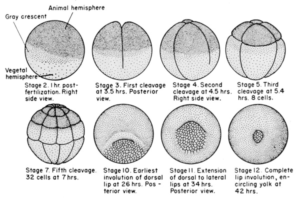
When sperm fertilize the egg, streaming movements are set up in the egg and these results in distribution of materials. So that three regions can be seen, the upper animal hemisphere (pole) which is pigmented and lower white vegetal pole. Between the two hemispheres, there is a small are with no pigment called grey crescent.
Cleavage or Segmentation:
Figure: stages of cleavage
2-3 hours after fertilization, the zygote begins to divide. The repeated division in the successive fashion is known as cleavage or segmentation.
Division is mitotic
The cleavage begins as a small depression at animal pole and gradually extends surrounding the zygote, dividing into two cell.
The divisions are holoblastic and complete
First cleavage is vertical; two celled stage
Second cleavage is also vertical but right angle to the first one; forms 4 celled stage
The cells are known as blastomere
Third cleavage is horizontal but above the equatorial line forming unequal size cells. The upper 4 cells toward animal pole are small and pigmented known as micromeres or epiblast. The lower 4 large yolk laden cells are known as megameres or hypoblat.
Fourth and fifth cleavage are also vertical forming 16 celled zygote. These division is followed by two horizontal cleacage, one toward animal pole and other toward vegetal pole, resulting in 32 celled stage.
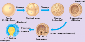
Morula (mulberry shape stage):
As the result of repeated and irregular cleavage, ball of cells is formed known as morula stage.
One hemisphere of morula is composed of large number of small black and yolkless cells known as micromeres and other hemisphere is composed of fewer number of large white and yolk laden cells known as megameres.
Blastula stage:
The micromeres dives more rapidly than megameres which results in formation of small fluid filled cavity known as Blastocoel or segmentation cavity.
Blastocoel bearing stage is called Blastula
The floor of blastocoel is composed of layer of yolk laden megameres while the roof is composed of micromeres.
In this stage early Presumptive areas can be differentiated by staining technique.
The entire animal pole of blastula represents the presumptive ectoderm, which is further divided into presumptive epidermis and presumptive neural plate
A small area near vegetal pole is presumptive notocord
Close to presumptive norocord there is a grey crescent region which is the presumptive mesoderm
The remaining vegetal region is presumptive endoderm
Gastrula stage:
Gastrula is the two layered embryo stage formed by migration and rearrangement of cells of blastula. The process of formation of gastrula is called gastrulation.
Gastrulation involves some critical changes in the blastula such as- differentiation of cells, transformation from monoblastic to diploblastic layer, formation of three primary germ layers.
Gastrulation completes in following steps. Figure: stages of gastrulation
Epiboly:
In this step, micromeres at animal pole dives more repeatedly and rapidly enclosing the megameres except in the region of yolk plug. This overgrowth or spreading of micromere cells is known as Epibloy.
2. Emboly or Intucking (Invagination):
In this step, small groove appears due to invagination of megameres near grey crescent region. The invagination gradually grows inward causing migration of cells.
This stage is also known as Yolk plug stage.
The narrowing of blastopore exerts pressure on underlying yolk laden megameres, result in protruding of some megameres cells as yolk plug.
Contraction of lips of blastopore: contraction of lips from all side occurs so that blastopore become smaller and narrower.
As invagination progresses archenteron increases in size and the blastocoel become reduced and finally obliterated.
This groove is the beginning of archenteron and its anterior opening is called blastopore. The blastopore is guided by anterior margin called dorsal lip and backward projecting lateral lip.
3. Involution:
due to increase in size of archenteron as well as formation of yolk plug, there is rapid migration of presumptive areas within the embryo occurs. This movement of the presumptive areas is known as involution.
Rotation of gastrula: gastrulation causes shift in the center of gravity of the embryo. In the blastula stage, embryo floats with animal pole upward. But formation of archenteron causes the embryo to rotate within the vitelline membrane so that blastopore comes near the vegetal pole.
Gastrulation causes following changes-
i) blastopore is presumptive gut
ii) roof of archenteron is chordamesoderm
iii) floor of archenteron is endoderm
4. Formation of three germ layer:
The three layers are ectoderm, mesoderm and endoderm are known as primary germ layer. They are also called as germinal layers because entire organs and body are derived from these layer.
Fate of germ layers
Ectoderm: epidermis, cutaneous glands, eye lens, cornea, retina, conjunctiva, central nervous system (brain and spinal cord), pineal gland, pituitary gland, enamel of teeth etc are derived from primary ectoderm layer.
Mesoderm: notochord, pericardium, peritoneum, muscles, skeleton, connective tissues-blood, lymph, adipose tissue, dermis of skin, visceral organs, are derived from primary mesoderm layer.
Endoderm: epithelium of digestive tract, respiratory tracts, Eustachian tubes, gastric and intestinal glands, liver, pancreas, bile and pancreatic ducts, lining of urinary bladder are derived from primary endoderm layer.
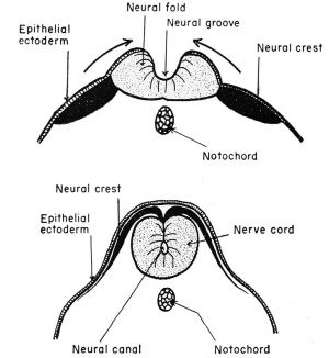
Neurulation:
Figure: process of formation of nerve cord
It is the process of formation of neural tube or nerve cord.
At the end of gastrulation the prospective neural plate comes to lie along the length of mid-dorsal region. Neural plate later forms central nervous system including brain and spinal cord.
A pair of longitudinal ridges called neural folds appears along the edges of neural plate, which meet in a semicircle anteriorly.
The neural folds increase in height and comes closer together the median line where they fuse to form neural tube, enclosing the neural canal.
The closure of neural tube begins just in front of mid-region and proceeds both anteriorly and posteriorly
At the front end neural tube remains open for short time through neuropore. But posteriorly it communicates for some time with archenteron by neurenteric canal.
Finally closed tubular neural tube is formed which later form brain and spinal cord.
Notogenesis:
It is the process of formation of notochord
The meso-endodermal cell lying in mid dorsal region of roof of archenteron separates from mesoderm layer
These cells become solid cylinder rod like structure along the median line and parallel to and just below the neural tube lies called notochord.
Later, notochordal sheath develop around the notochord
In adult notochord is replaced by vertebral column.
Formation of coelom:
Coelom is the body cavity and it is mesodermal in origin
Mesodermal layer split into two thin layers-outer somatic or (parietal) layer and inner visceral or (splanchnic) layer.
Between these two layers a cavity is formed called splanchnocoel, which extend downward and continues to the outside below the gut
Outer somatic layer combines with ectoderm to form body wall (somatopleure)
Inner visceral layer unites with endoderm to form gut wall (splanchnopleure)
Splanchnocoel continues to form coelom or body cavity between gut wall and body wall.
The coelom is known as Schizocoel coelom.
Developmental biology of Frog-Embryonic Development