
Circulatory system of the earthworm:
- In earthworm, the blood vascular system is of closed type.
- Blood vascular system is different in first 13 segments as regards to number, arrangements and nature of blood vessels.
- It comprises of the blood vessels, hearts, loops, blood capillaries and the blood glands.
Blood of earthworm:
- The respiratory pigment haemoglobin is dissolved in plasma, and hence the blood of the earthworm is red in colour.
- The plasma also includes other corpuscles that are colourless and bear nucleus.
Blood Vessels of earthworm:
- The blood vessels are of two distinct types.
- They can either be collecting blood vessels or distributing blood vessels.
- The vessels are closed tubes and possess definite wall.
- They further break down into capillaries and are divaricated in the various parts of the body.
- The arrangement of blood vessels in the anterior thirteen segments slightly differs from that behind the thirteen segment, i.e., in the region of intestine.
- Thus, the simplified headings for the study of blood vessels are:
- Blood vessels and its arrangement in the segments behind 13th, i.e., intestinal region.
- Blood vessels and its arrangement in the anterior thirteen segments.
A] Blood Vessels behind 13th segments in Intestinal Region:
- The blood vessels of intestinal region comprises of:
- Median longitudinal blood vessels
- The intestinal blood plexus
- The commissural vessel
- The integumentary vessel
- The nephridial vessels
1. Median Longitudinal Blood Vessels:
- (i) Dorsal blood Vessel:
- It is located above the intestine in mid dorsal line.
- The direction of flow of blood in dorsal vessel is from posterior to anterior.
- Dorsal blood vessel is the largest blood vessel of the body in earthworm.
- It is the thickest blood vessel having contractile muscular walls.
- It is visible as a dark line from the thin and semi-transparent body wall.
- It is contractile and rhythmically operates to force the blood from the posterior to the anterior side.
- It has a pair of valves internally in each segment which check the backward flow of blood.
- Dorsal blood vessel is regarded as true heart in earthworm.
- Dorsal blood vessel is distributive in segments 1-13 and main collecting vessel in the segments 14 onwards.
- From the posterior segment up to the 14th segment, It collects blood from 2 pairs of dorso-intestinal vessels from the intestine in each segment and a pair of commissural vessels from the sub-neural vessel.
- Behind each septum, the commissural vessels create a loop and receive blood from the body wall, nephridia and prostate glands.
- Through a septo-intestinal branch, the commissural vessels distribute the blood in each segment into the intestine.
- (ii) Ventral blood Vessel:
- Ventral blood vessel is found below the alimentary canal and above the ventral nerve cord.
- It is also a long blood vessel that runs from second segment to the last segment of the body.
- It is the main distributive blood vessel of earthworm.
- It has thin wall and lacks muscles and valves.
- The direction of the flow of blood is anterior to posterior side.
- In the anterior segments (i.e. first 13 segments), ventral blood vessel supplies blood to the body wall, septa, nephridia, and reproductive organs of the same segment through the ventro-tegumentaries (a pair in each segment).
- Ventral blood vessel supplies blood to the intestine in the intestinal region through the single median ventro-intestinal in each segment.
- Similarly, it gives off a pair of ventro-tegumentaries, one on each side, in front of septum in each segment.
- This ventro-tegumentaries supply blood to the body wall, septa and integumentary nephridia, septal nephridia, gonads, spermathecae and seminal vesicles.
- Behind the 13th segment, the ventral vessel also branch out a ventro-intestinal vessel in each segment.
- These carry blood to the lower part of the intestine.
- The blood plexuses are formed by the branches in intestine consisting of two networks in the intestinal wall.
- (iii) Sub-neural Vessel:
- It is long and thin vessel and is situated mid-ventrally below the ventral nerve cord.
- Sub-neural vessel runs from 14th segment up to last segment, below the ventral nerve cord.
- It lacks muscular walls and internal valves.
- It is mainly a collecting blood vessel.
- The direction of blood flow is from anterior to posterior end.
- It receives a pair of slender branches in each segment which bring blood from the ventral body wall and the nerve cord.
- It branch off a pair of commissural vessels in each segment which join the dorsal vessel.
- Hence, it receives blood from the ventral body wall and supplies some blood to the intestine.
2. Intestinal Blood Plexus:
- The blood capillaries supply the intestine of earthworm that forms a close network.
- The intestinal blood plexus is comprised of a close network of capillaries in the wall of intestine.
- Two capillary networks are present in the intestine i.e. (i) External and (ii)Internal
- The capillary network found at the outer surface of intestine is termed as external plexus.
- External plexus receives blood from the ventral vessel through ventro-intestinal and passes it on to the internal plexus.
- The capillary network found between the circular muscle layer of intestine and its internal epithelial lining is referred as internal plexus.
- Internal plexus is linked with dorsal blood vessel through the dorso-intestinals.
- Internal plexus functions to absorb the nutrients from the gut.
3. Commissural Vessels:
- Commissural vessels link the dorsal and sub-neural vessels.
- These vessels receive blood from nephridia, body wall and reproductive organs through capillaries.
- Then, they supply it to dorsal blood vessel.
4. Integumentary Vessels:
- These vessels originate from ventral vessels.
- It supplies the blood to integument for aeration and the aerated blood is collected by various capillaries of commissural vessel in each segment.
- Hence, there exists an intimate parallelism among venous and arterial capillaries throughout the body wall.
5. Nephridial Vessels:
- Nephridial vessels arises from the ventro-tegumentary vessels of ventral vessel.
- It supplies the blood to the nephridia.
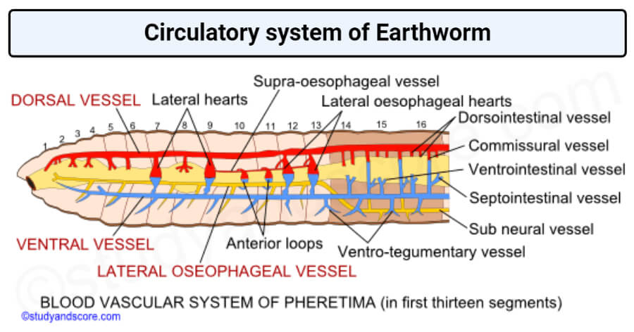
B] Blood Vessels anterior to 13 Segments:
- The blood vascular system in the first thirteen segments is modified considerably and differs markedly from that of the intestinal region.
- It comprises of :
- Median longitudinal vessels;
- Hearts and anterior loops;
- Blood vessels of the gut.
- The function of collecting blood from the anterior region of the gut is taken over by a new vessel supra-oesophageal, while the blood from the peripheral structures is collected by the right and left lateral oesophageal.
1. Median Longitudinal Blood Vessels:
- (i) Dorsal blood vessel:
- In anterior 13 segments, dorsal blood vessel becomes distributive in function.
- It is similar in structure as in posterior segments, however it lacks dorso-intestinalis and commissural vessels opening into it.
- It exports all the collected blood from the posterior region of the body into hearts and the anterior region of the gut.
- Here, it gives off three branches that is distributed over the pharyngeal bulb and the roof of the buccal chamber.
- However, it supplies blood to stomach, gizzard, oesophagus, pharynx and other related parts.
- (ii) Ventral blood vessel:
- Ventral blood vessel is again distributve in these segments as well.
- However, it extends only up to the second segment.
- Due to the absence of ventrointestinals, it does not supply to the alimentary canal in this region.
- In the anterior segments (i.e. 1st 13 segments), ventral blood vessel supplies blood to the body wall, septa, nephridia, and reproductive organs of the same segment through the ventro-tegumentaries (a pair in each segment).
- (iii) Supra-oesophageal vessel:
- It is smallest vessel of body lying from 9th to 13th segment.
- It is located above the stomach.
- It is the main transverse vessels of first 13 segments.
- It gets blood from the lateral oesophageals through two pairs of anterior loops that surround the stomach in the 10th and 11th segments.
- It carries collected blood by the latero-oesophageal hearts in 12th and 13th segments to the ventral vessel.
- (iv) Lateral oesophageals:
- In the 14th segment, the sub-neural vessel actually bifurcates to form two lateral oesophageals.
- In the anterior thirteen segments, these vessels are comparatively thick and located along the ventro-lateral margins of the alimentary canal.
- These vessels are tightly connected to the wall of the stomach from the 10th to 13th segments and interact with the ring vessels.
- However, in the gizzard region and further forwards, they exist free from the wall of the alimentary canal.
- They continue receiving branches from it in each segment.
- In each segment, these vessels receive a pair of branches that hold blood from the body wall and the septum.
- They also collect blood from the reproductive organs and nephridia, thus acting like the posterior region’s sub-neural and commissural vessels, i.e. these are vessels that collect.
- It collects blood from seminal vesicles present in 11th and 12th segments.
2. Hearts and Anterior Loops:
- The dorsal and ventral blood vessels have no direct connections in the posterior segments behind 13th segment.
- However, in the anterior region both these vessels are connected together by 4 pairs of pulsatile hearts.
- The hearts are neurogenic i.e. the heart beat arises in the nerve cells of the heart.
- The hearts are contractile and surround the alimentary canal, they are present in the segments 7th , 9th , 12th and 13th.They can further be simplified as:
- Lateral hearts:
- One pair of lateral hearts in 7th and one pair in 9th segment.
- Each lateral heart possesses 4 pairs of valves that permits blood to flow downwards only.
- It sends blood from dorsal vessel to ventral vessel.
- Lateral oesophageal hearts:
- One pair lateral oesophageal hearts in 12th and one pair in 13th segment.
- Each lateral oesophageal heart has thick muscular walls.
- It possesses 3 pairs of valves and sends blood from supraoesophageal and dorsal vessel to ventral vessel.
- A pair of valves is present at each junction with the dorsal vessels and supra-oesophageal vessel, and remaining pair of valves at the ventral end.
- These valves allow flow of blood to downwards only.
- Anterior loops:
- Along with four pairs of hearts, there are two pairs of loop-like vessels that connects the supra-oesophageal with the lateral oesophageals.
- These vessels are neither muscular nor pulsatile and are termed as anterior loops.
- These lack valves.
- One pair is present in 10th and one pair in 11th segment.
- It sends blood from lateral oesophageal vessel to supraoesophageal vessel into ventral vessel through the hearts of 12th and 13th segments.
3. Blood Vessels of the Gut:
- These are ring-like vessels that connect the supra-oesophageal and lateral-oesophageal vessels.
- These are located on either side of stomach.
- Through these vessels blood flows upwards from the lateral- oesophageals into the supra-oesophageal.
- Dorsal blood vessel supplies blood to buccal cavity, pharynx and gizzard directly.
How is blood circulated in earthworm?
- The blood collected by the dorsal vessel through the dorsointestinals, intestinal blood plexuses, and commissurals is distributed partly to the anterior alimentary canal, but primarily to the ventral vessel through the heart.
- The blood flows forward to the anterior region in front of the hearts in the ventral vessel.
- However, the largest proportion of blood flows backwards, which is distributed to the body wall and the organs in the coelom via ventrotegumentaries and to the alimentary canal via the ventrointestinal vessels.
- In other terms, ventral vessel supplies blood to all parts.
- The sub-neural collects blood from the ventral body wall, which also receives some blood from the anterior region via the lateral-oesophageal region.
- This blood passes from the sub-neural to the dorsal vessel via the commissurals.
- The lateral oesophageals also send blood to the supra-oesophageal vessel through the anterior loops, which then passes it to the ventral vessel through the latero-oesophageal heart.
What are the functions of blood in earthworm?
- The blood distributes digested food to different regions of the body and absorbs waste materials such as nitrogen waste and CO2 that are administered to nephridia, skin and coelomic fluid.
- Respiration takes place in almost all aquatic and terrestrial oligochaetes by the diffusion of gases through the integument, which comprises a capillary network in the outer epidermal layer in larger forms.
- The film of moisture needed for the diffusion of gases is provided by mucous glands, coelomic fluid, and nephridial excretions.
- Plasma haemoglobin absorbs O2 from the capillaries of the skin, but moist skin must be available where O2 can be mixed with haemoglobin in order to be carried by blood.
- Haemoglobin is an appropriate pigment and can absorb O2 either from the ambient air or from a relatively oxygen deficient environment.
- Earthworms can also survive in well-aerated water and do not drown.
- They can also survive without O2 for several hours, potentially carrying on anaerobic respiration in this state.
Blood Glands in Earthworm:
- Blood glands are various groups of small rounded follicles of red colour.
- The follicles have a syncytial wall surrounding a capsule containing a mass of loose cells.
- The blood glands are present in the 4th, 5th, and 6th segments of Pheretima above the pharyngeal mass.
- Blood glands synthesizes blood corpuscles and hemoglobin are synthesized.
- Blood glands are related with pharyngeal nephridia and with salivary glands.
- Hence, these glands may be excretory as well.
Lymph glands in Earthworm:
- On both sides of dorsal blood vessel from 26th segment and those behind it (one pair per segment, small and whitish), lymph glands are found.
- Lymph glands are responsible for producing certain phagocytic cells.
Summary points on Earthworm circulatory system:
- In earthworm, the blood vascular system is of closed type.
- Blood vascular system is different in first 13 segments as regards to number, arrangements and nature of blood vessels.
- Dorsal blood vessel is distributive in segments 1-13 and main collecting vessel in the segments 14 onwards.
- Ventral blood vessel is the main distributive blood vessel of earthworm.
- Supra oesophageal is smallest vessel of body lying from 9th to 13th segment.
- Lateral oesophageals collects blood from seminal vesicles present in 11th and 12th segments.
- One pair of anterior loops is present in 10th and one pair in 11th segment.
- Dorsal blood vessel supplies blood to buccal cavity, pharynx and gizzard directly.
- The blood glands are present in the 4th, 5th, and 6th segments of Pheretima above the pharyngeal mass.