Chromosome banding technique:
- Study of chromosome number and structure by staining the dividing cells with certain dyes and then examining them under microscope for cytogenetic analysis is called chromosome banding.
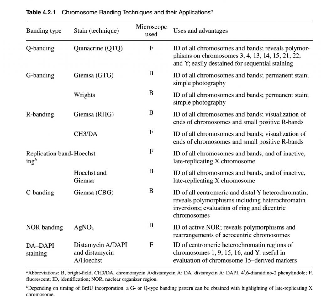
- Most cytological analysis is performed on dividing cells (metaphase of mitosis). Therefore, meristematic cell of root or shoot tip of plant and embryo cell of animals are used. However, the development of cell-culturing techniques has made it possible to study chromosomes in other types of cells. For example, human white blood cells can be collected from peripheral blood, separated from the non-dividing red blood cells, and put into culture. The white cells are then stimulated to divide by chemical treatment, and midway through division a sample of the cells is prepared for cytological analysis.
- The dividing cell on treatment when treated with chemical disables spindle fibre formation, such mitotically arrested cell are then immersed in a hypotonic solution that causes cell to take up water. On preparation for microscopic examination such cells are squashed on a microscopic slide, such that the chromosomes are spread out in an uncluttered fashion which can be observed by staining
- The stains such as Feulgen’s reagent or aceto-carmine stains the chromosome uniformly making difficult to distinguish one chromosome form other. Now a days, cytogeneticists use dyes that stain chromosomes differentially along their lengths.
- The staining of chromosome is known as banding technique because stains give rise to pattern of bands along the length of chromosome.
Types of chromosome banding technique:
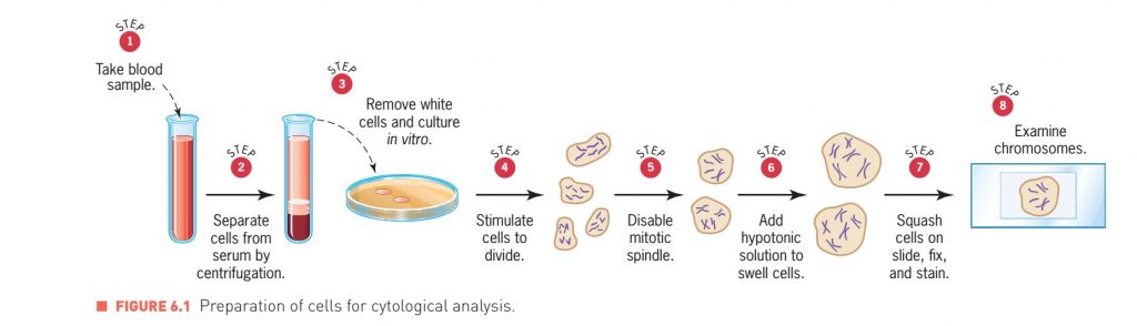
1. Q-banding:
- Q banding used quinacrine stain (quinacrine dihydrochloride or quinacrine mustard) and it is the simplest and the first chromosomal banding method. Quinacrine stained chromosomes show a characteristic pattern of bright bands on a darker background. Because quinacrine is a DNA intercalating agent and a fluorescent compound, the bands appear only when the chromosomes are exposed to ultraviolet (UV) light.
- Ultraviolet light causes the quinacrine molecules to shine, therefore parts of the chromosome intercalated with quinacrine shine brightly, whereas other parts remain dark.
- This bright-dark banding pattern is highly reproducible and is also specific for each chromosome. Thus with quinacrine banding is used to identify particular chromosomes in a cell, and also to determine if a chromosome is structurally abnormal.
- Quinacrine is an intercalating agent and it intercalate between the base pairs of the DNA helix. Quinacrine has more affinity for DNA sequence containing AT sequence. Therefore fluorescence of quinacrine is enhanced along AT rich sequence and appear bright than GC rich sequence.
- * quinacrine is carcinogenic.
2. G-banding:
- G-banding is the most frequent used technique for chromosome staining in cytogenetics.
- Giemsa staining is an excellent nonfluorescent staining techniques.
- Giemsa also creates a reproducible pattern of bands on each chromosome. It is still not clear why chromosomes show bands when they are stained with quinacrine or Giemsa
- Bright field microscope is used for visualization
- In G banding technique, before using Giemsa stain, there is always a pretreatment step.
- Usually proteolytic enzyme trypsin is used for pretreatment. Therefore the process is also known as GTG banding (G-banding by trypsin with Giemsa).
- There is an alternative to Giemsa stain and it is Wright stain.
- G-banding also produce same banding pattern as Q banding along the length of chromosome. Geimsa stain has more affinity for DNA sequence rich in AT content hence stained dark while sequence rich in GC content stain light.
3. C-banding:
- C-banding is called Centromeric heterochromatin staining
- In this technique, before using Giemsa stain the cell is pretreated with alkali. Theefore this technique is also known as CBG-staining (B-banding by base with Giemsa).
- Centromeric heterochromatin and distal part of Y chromosome containing highly repetitive DNA sequence (satellite DNA) is used to stain by C-binding technique.
- After staining, banding is viewed by bright-field microscope.
4. R-banding:
- R-banding is called Reverse chromosome banding.
- With this banding technique the band pattern produced in chromosome is reversed to the band produced by G-banding and Q-banding. ie. The dark band (AT rich region) observed in G-banding technique appears light in R-banding and vice versa.
- R-banding technique also uses Giemsa stain but before staining with Giemsa the slide is heated at 88°C in a buffer solution. Heating causes denaturation of DNA.
- Denaturation of chromosome at AT rich region occurs at faster rate resulting in loss of DNA from these regions but not from GC rich region. The GC rich region is then stained by Giemsa stain which appears stained (R band)
- G-banding is usually preferred over R-banding. However R-banding can be used for chromosome identification.
- In some cases, R-banding is a useful complement to G-banding because some small light G band can be more easily detected when they are stained by R-banding.
- R-banding is also useful for visualization of telomere sequence at the ends of chromosomes. Telomeres stained dark with R-banding while light with G-banding.
Chromosome painting:
- It is the most advanced technique. With this technique, colorful chromosome images are created by treating chromosome spreads with fluorescently labeled DNA fragments that have been isolated and characterized in the laboratory.
- At first the DNA fragment isolated from desired gene is labelled with florescent dye and is applied to chromosomal spread on a slide. The florescent labelled DNA fragment will bind to chromosomal DNA that is complementary to it in sequence. This binding results in visual color at the position of complementary sequence.