
Introduction and Epidemiology of Brugia malayi:
- Brugia malayi is a filarial worm belongs to phylum nematoda which is one of three causative agents of elephantiasis (lymphatic filariasis) in humans. ( other are- Wuchereria bancrofti, Brugia timori)
- Brug in 1927 describe for the first time about new type of microfilaria in blood of natives in Sumatra. From where the genus acquired its name, Brugia.
- The adult B.malayi worm of was described by Rao and Maplestone in India (1940).
- B. malayi has a more restricted distributions than other filarial worms and is commonly found in open swamps and the rice growing areas of coastal regions.
- It is endemic in India, Srilanka, Philippines, Southern Thailand, North Veitnam, China, South Korea and Japan.
- Besides B. malayi, the genus also includes B. timori, which is human parasite causing lymphatic filariasis.
- Other animal species such as B. pahangi and B. patei infecting dogs and cats.
- Human are the primary host.
- Leaf monkeys are also definitive hosts and reservoir of sub periodic B. malayi. Hence, zoonotic infection can occur from infected monkeys to humans.
- Mosquitoes are the intermediate host.
Habitat
- The adult worm B. malayi is found in the lymphatic system of man and other mammals.
- Microfilaria are mainly found in the peripheral blood circulation.
Morphology of Brugia malayi:
Adult worm:
- The adult worm is similar to Wuchereria bancrofti but are smaller in size.
- The worm resembles a delicate white thread.
- The mature females vary in length from 4.3 – 5.5 cm and in breadth from 0.13-0.17 mm.
- Mature males measure 1.2-2.3 cm in length and 0.07- 0.08 mm in breadth.
Microfilaria
- Microfilaria are colorless and transparent with blunt heads and pointed tails in unstained preparations.
- They lie folded with head close to tail.
- Each microfilaria measures 177-230 µm in length and 5-6 µm in diameter. They possess secondary kinks, instead of smooth curves. The presence of two distinct nuclei at the tip of the tail is the distinguishing feature of microfilaria.
- The nuclei is present at the extreme tip of the tail and other midway between the tip and the posterior column nuclei.
Third stage larvae: an Infective form
- L3 larva is the infective form of parasite are found in the mosquito vector.
- They are elongated, filariform and measure 1500 µm to 1800 µm in length and 18-23 µm in breadth.
Periodicity of Brugia malayi:
- B. malayi generally shows nocturnal periodicity. However, two variants of Brugia malayi are recognized.
- Nocturnal periodic Brugia malayi
- It is the most prevalent form of parasite
- Microfilaria show nocturnal periodicity
- Transmission occurs mainly by Anopheles and Mansonia mosquito
2. Sub periodic Brugia malayi
- It is less common form
- Microfilaria are found in the day time
- Transmission occurs by Mansonia and Coquillettidia mosquitoes
Pathogenesis of Brugia malayi
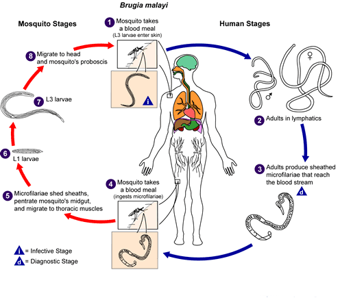
- Humans are the definitive host and mosquitoes are the intermediate hosts of Brugia spp. Infected human are the main sources and reservoir of infection. Man to Man transmission occurs by the bite of Anapheles and Mansonia.
- The life cycle of filarial parasites involves four larval stages and an adult stage.
- Infection begins with the bite of infected mosquito on the skin and deposition of infective stage larvae (L3).
- The larvae then pass through the puncture wound and reach the lymphatic system.
- Within the lymphatics and lymph nodes, the L3 larvae undergo molting and development to form L4 larvae.
- This takes about 7-10 days for both B. malayi and W. bancrofti
- The L4 larvae undergo a subsequent molting/developmental step to form adult worms. This occurs about 4-6 weeks after L3 entry in the case of B. malayi.
- The adult worms take permanent residence in afferent lymphatics or the cortical sinuses of lymph nodes and generate microscopic live progeny called “microfilariae”.
- The female worms can give birth to as many as 50,000 microfilariae per day, which find their way into the blood circulation from the lymphatics.
- The adult worms are estimated to survive for a period of 5-10 years although longer durations have been recorded.
- The microfilariae of B. malayi, for the large part, exhibit a phenomenon called nocturnal periodicity, i.e., they appear in larger numbers in the peripheral circulation at night and retreat during the day.
- Subperiodic or nonperiodic of B. malayi are also found in certain parts of the world.
I. Pathology of acute filariasis
Lymphadenitis:
- It is the typical feature. The more classical is acute filarial adenolymphangitis, which is felt to reflect an immune-mediated inflammatory response to dead or dying adult worms.
- The attacks of lymphadenitis occur at regular intervals and often are precipitated by hard muscular exercise.
- The episodes of attacks vary from 1-2 attacks per year.
- Lymphadenitis typically occurs in the inguinal region.
- Occasionally the auxillary lymph nodes are also involved.
- It occurs infrequently at atypical sites such as the popliteal lymph nodes or breasts.
Lymphangitis
- Lymphadenitis is followed by Retrograde lymphangitis
Lymphatic abscess
- The infected lymph nodes may suppurate and form abscess.
- These are usually superficial.
- They rupture leaving behind ulcers.
II. Pathology of chronic filariasis
- The common sites of elephantiasis include, the leg below the knee and less frequently the arm below the elbow.
- Genital involvement and chyluria characteristically are absent.
Clinical manifestation of lymphatic filariasis:
- Incubation period is short and varies from 6-16 month.
- B. malayi shows following clinical stages.
i. Endemic normal and asymptomatic stage:
- Most infection are asymptomatic and common among infected persons in endemic areas with no symptoms of filarial infection and yet, on routine blood examinations, demonstrate the presence of significant numbers of parasites.
ii. Acute filariasis:
- It is characterized by recurrent attacks of lymphadenitis (inflammation of lymph nodes) and lymphangitis associated with fever, chill and other constitutional symptoms.
- The attacks are followed by a characteristics retrograde lymphangitis. The affected lymph vessels become cord-like and tender. The frequency of attacks per year to several attacks per month.
- The infected lymph nodes may suppurate and form abscesses.
- The lymph node abscess is the characteristics of Malayan filariasis.
- Incomplete resolution of the oedema after each attacks leads to the characteristics chronic stage.
Chronic filariasis:
- It is characterized by elephantiasis that develops after 10-15 years in a small number infected population.
- The legs below the knee, less frequently the arms below the elbow are characteristically affected.
- Genital involvement and chyluna are not reported in Malayan filariasis, this occurs only along with bancroftian filariasis.
Lab Diagnosis
- Same as lab diagnosis of W. bancrofti
Treatment of Brugia malayi
- Diethylcarbamagine (DEC): is relatively effective in smaller dose.
- Dose regimen consists of 50 mg on first day; 50 mg three times daily on 2nd day; 100 mg three times daily on 3rd day and finally 2 mg/kg/day in three divided doses from 4th day till 21st day.