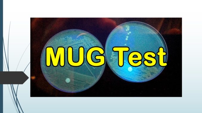
Principle of MUG test:
- About 97% strains of Escherichia coli and organisms as such Salmonella, Shigella, and Yersinia are capable of producing enzyme β-glucuronidase. MUG stands for 4-Methylumbelliferyl-β-D Glucuronide which is a substrate hydrolysed by the enzyme β-glucuronidase. During this hydrolysis, 4-methylumbilliferone is released which fluoresces under long wave UV light which is visible as blue colour. This MUG test is performed for rapid identification of E. coli in the clinical specimens. Since, verotoxin producing strains of E. coli are incapable for producing MUG thus, MUG test can be useful for the determination of such strains due to absence of the enzyme in the sample. The fluorescence if visible indicates the breakdown of substrate and is regarded as positive test, and the negative test is indicated by no fluorescence suggesting that no any reaction occurred due to absence of enzyme.
Requirements:
- MUG disk
- 2-3 isolated colonies of organisms
- Inoculating loops
- Petri dishes and test tubes
- Incubator
- Long wave UV light
Procedure of MUG test:
i. Direct disk test:
- Place a MUG disk in an sterile petri dish and add a drop of distilled water.
- Streak out 2-3 isolated colonies from 18-24 hour old culture on the disk.
- Place a piece of filter paper saturated with water in the lid of the petri dish to maintain humid environment.
- Incubate aerobically at 35°-37°C for 30 minutes.
- Following incubation, examine the disk for fluorescence using longwave ultraviolet light (360nm) in a darkened room.
ii. Tube test:
- Insert 0.25ml of deionized water to a clean glass tube.
- Create a heavy suspension with 3- 4 colonies of test isolate in the tube.
- By use of forceps, place a MUG disk in the tube and shake strenuously to make sure the adequate elution of substrate in the surrounding liquid.
- Incubate aerobically at 35°-37°C for 1 hour.
- Following incubation, examine the disk for fluorescence by use of longwave ultraviolet light (360nm) in a darkened room.|
Results interpretation:
- Positive result: The blue fluorescence is suggestive of positive test.
- Negative result: Absence of blue fluorescence is indicative of negative test.

Limitations:
- Since not all E. coli are MUG positive, we cannot conclude the organism tested is not E. coli even if it gives negative test.
- Media that contains dyes should be avoided.
- Oxidase positive organisms as such Pseudomonas aeruginosa gives fluorescence and may resemble MUG test, thus this test should not be performed in oxidase positive organisms.
- Orange fluorescence by some organisms should be considered as negative test.
- Serological testing maybe required for differentiating Shigella and E. coli as some strains of Shigella are MUG positive.
