
Characteristics of Bacteroides
- Gram negative anaerobic rod
- Shape: Pleuromorphic
- Size: (0.5-1.5)µm wide and (2-6)µm long
- Non motile except B. polypragmatus, B. xylanolyticus
- Non capsulated except fragilis
- Non spore forming
- Habitat: Norma flora of gastrointestinal tract, mouth, skin, nasopharyns, Upper respiratory tract, vagina
- Opportunistic human pathogen
Classification of Bacteroides
- on the basis of medical importance bacteriodes is classified into two group
1. Bacteroides fragilis group:
- Examples:
- B.fragilis
- B. distasonis
- B. ovatus,
- B. thetaiotaomicron
- B. vulgatus
- B. idgatus
- B. uniformis
- B. tiariabilisleggerthii
- B. splanchnicus
- these are commensals of GI tract
2. Bacteroides melaninogenicus group
- Examples:
- B. melaninogenicus sub spp intermedius
- B. ruminicola
- Now the name has changed to Prevotella melaningenica
- these are Norma flora of URT, GIT, vagina
Pathogenesis:
1. Mode of transmission:
- Displacement of their normal habitat
2. Virulence factors of Bacteroides
- i) Capsule:- Helps in attachment, and resist phagocytosis and complement mediated lysis.
- ii) Fimbriae: helps in attachment
- iii) Endotoxin (LPS):- stimulates leucocytes
- iv) Short chain Fatty acids (succinic acid):- resist phagocytosis and intracellular killing
- v) Enzymes- protect from Oxygen toxicity
- Catalase and Superoxide dismutase: These enzymes inactivates H2O2 and release super oxide free radicals there by protect anaerobes when exposed to oxygen
- Protease
- Collagenase
- Phospholipase
- Neuraminidase
- Heparinase
- Haemolysin
- Fibrinolysin
- Gluconidase
Clinical manifestation of Bacteroides:
- Genitourinary tract infection
- Appendicitis
- Bacteriaemia
- Endocarditis, pericarditis, vascular graft infection
- Meningitis
- Septic artheritis
- Bed sore
- Inflammation of viscent angina ( throat)
- Skin and soft tissue infection
- Upper respiratory infection
Laboratory diagnosis of Bacteroides
Samples: pus, exudates, biopsy
1. Microscopy:
- gram negative, non motile, non sporing pleuromorphic rod
2. Culture:
i) Blood Agar (BA):
- Kanamycin or neomycin Blood agar selective for Anaerobes
- Inculates anaerobically at 37C for 48 hours
- fragilis Form non haemolytic grey colony of 1-3 mm diameter
- melanogenicus form black brown haemolytic colony in 3-5 days
ii) Bactrroides Bile Esculin Agar (BBE):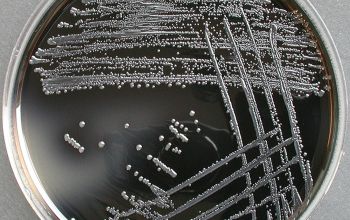
- hydrolyses esculin,
- colony surrounded by dark zone
3. Biochemical tests for fragilis
- i) Esculin hydrolyses: positive (+)
- ii) Indole: negative (-)
- iii) Urease: Negative (-)
- iv) Catalase: Positive (+)
- v) Oxidase : variable (+/-)
- vi) Fermentative: glucose +, lactose +, maltose +, sucose + rhamnose -, Arabinose -, salicin -, trehalose –
- vii) Bile resistant: grow in 20% bile
- viii) Thioglycolate with bile: Positive (+)
- ix) Beta lactamase production: fragilis is resistant to penicillin
4. Serology
5. PCR
Treatment of Bacteroides:
- Metronidazole: drug of choice
- Cephalosporin
- Chloramphenicol