Sliding Filament Theory of Muscle Contraction
The mechanism of muscle contraction is explained by sliding filament model. This theory was proposed by H.E Huxley and J. Hanson, and A. F. Huxley and R. Niedergerke in 1954.
The arrangement of actin and myosin myofilament within a sarcomere is crucial in the mechanism of muscle contraction. It is proposed that muscle contracts by the actin and myosin filaments sliding past each other. For analogy, muscle contraction by sliding filament model is equivalent to interlocking fingers, pushing them together shortens the distance.
As sarcomere is the unit of muscle contraction, its length contracts resulting in whole muscle contraction. During contraction, length of A-band (Dark band) remains same whereas length of I-band (Light band) and H-zone gets shorter.
Actin myofilament:
- An actin myofilament is made up of actin molecule, tropomyosin and troponin complex. Troponin is composed of three sub-units (troponin I, T and C). Tropomyosin form two helical strand which are wrapped around actin molecules (G-actins) longitudinally in thin twisted stranded form.
- Each G-actin is attached with an ATP molecule. The whole assembly of actin molecules is known as F-actin (Fibrous actin).
- Tropomyosin switches ON or OFF the muscle contraction mechanism.
- Troponin complex is a globular protein which binds to tropomyosin and calcium ions.
Myosin myofilament:
- A myosin myofilament consists of two distinct region, a long rod-shaped tail called myosin rod and two globular intertwined myosin head.
- The globular head appear at interval along the myosin myofilament, projecting from the sides of the filament.
- The myosin head can attach to the neighboring acting filament where actin and myosin filaments overlaps.
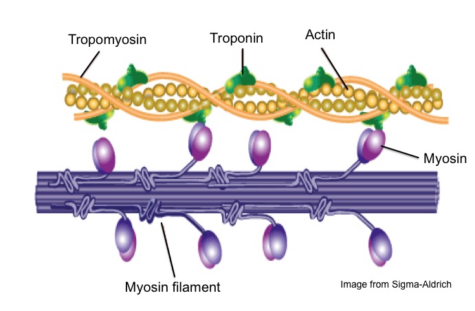
Figure: Actin and Myosin myofilament
Mechanism of Muscle contraction:
- When the nerve impulse from brain and spinal cord are carried along motor neuron to neuromuscular junction, Ca++ ions are released in the terminal axon. Increases calcium ion concentration stimulates the release of neurotransmitter (Acetylcholine) in the synaptic cleft. The neurotransmitter binds to the receptor on the sarcolemma and depolarization and generate action potential across muscle fiber for muscle contraction. The action potential propagates over entire muscle fiber and move to the adjacent fibers along transverse tubules. The action potential in transverse tubules causes the release of calcium ion from sarcoplasmic reticulum, which stimulate for muscle contraction.
The sequences of muscle contraction explained by sliding filament theory are as follows
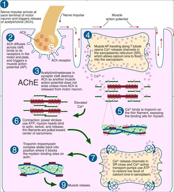
Figure: diagrammatic representation of muscle contraction mechanism
1. Blocking of myosin head:
-
Actin and myosin overlaps each other forming cross bridge. The cross bridge is active only when myosin head attached like hook to the actin filament.
-
When muscle is at rest, the overlapping of actin filament to the myosin head is blocked by tropomyosin. The actin myofilament is said to be in OFF position
2. Release of calcium ions:
-
Nerve impulse causing depolarization and action potential in the sarcolemma trigger the release of calcium ions from sarcoplasmic reticulum.
-
The calcium ion then binds with the troponin complex on the actin myofilament causing displacement of troponin complex and tropomyosin from its blocking site exposing myosin binding site.
-
As soon as the myosin binding site is exposed, myosin head cross bridge with actin filament. Now, the actin myofilament is said to be in ON position.
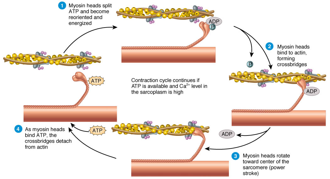
3. Active Cross-bridge formation:
-
When myosin head attached like hooks to the neighboring actin filament, active cross bridge is formed. The cross bridge between actin and myosin filament acts as an enzyme (Myosin ATPase).
-
The enzyme Myosin ATpase hydrolyses ATP stored into ADP and inorganic phosphate and release energy. This released energy is used for movement of myosin head toward actin filament. The myosin head tilts and pull actin filament along so that myosin and actin filament slide each other. The opposite end of actin myofilament within a sarcomere move toward each other, resulting in muscle contraction.
-
After sliding the cross bridge detached and the actin and myosin filament come back to original position. The active cross bridge form and reform for 50-100 time within a second using ATP in rapid fashion. Therefore, muscle fiber consists of numerous mitochondria.
-
In muscle contraction, sarcomere can contracts by 30-60% of its length