
Physiological events of vision consists of following;
- Refraction of light entering the eye
- Focusing of image on the retina by accommodation of lens
- Convergence of image
- Photo-chemical activity in retina and conversion into neural impulse
- Processing in brain and perception
Refraction of light entering the eye:
- Light wave travels parallel to each other but they bend when passes from one medium to another. This phenomenon is called refraction.
- Before light reach retina it passes through cornea, aqueous humor, lens vitrous humor, so refraction takes place in every medium before it falls on retina.
- In normal eye, light wave focused on retina.
- However in myopic eye (short sightedness) light focused in front of retina. So this defect can be treated by using cancave lens.
- In case of far sightedness light focused behind retina, so no image is formed. This defect can be treated by using convex lens.
Accommodation of lens to focus image:
- Accommodation is a reflex process to bring light rays from object into perfect focus on retina by adjusting the lens.
- When an object lying less than 6 meter away is viewed, image formed behind retina. But due to accommodation of lens image formed in retina and we can see the object.
- For accommodation to view closer object, ciliary muscle contract and lens become thick which causes focus on closer object.
- Similarly, when distant object is viewed, ciliary muscles relaxes, so the tension of ligament become greater which pull lens and lens become thinner, due to which image forms on retina.
- The normal eye is able to accommodate light from object about 25 cm to infinity.
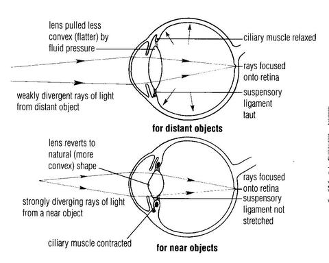
Focus on nearer object:
Ciliary muscle contract——-ciliary body pull forward and inward ———tension on suspensory ligament of lens reduced ——lens become thicker and round due to its elasticity ——possible to focus near object
Focus on distant object:
Ciliary muscles relaxes——ciliary body return to its normal resting state—–tension on suspensory ligament of lens increases——-lens become thinner and flat———possible to focus distant object
Convergence of image:
- Human eye have binocular vision, it means although we have two eye, we perceive single image
- In binocular vision, two eye ball turns slightly inward to focus a close object so that both image falls on corresponding points on retina at same time. This phenomenon is called convergence.
Photo-chemical activity in retina and conversion into neural impulse
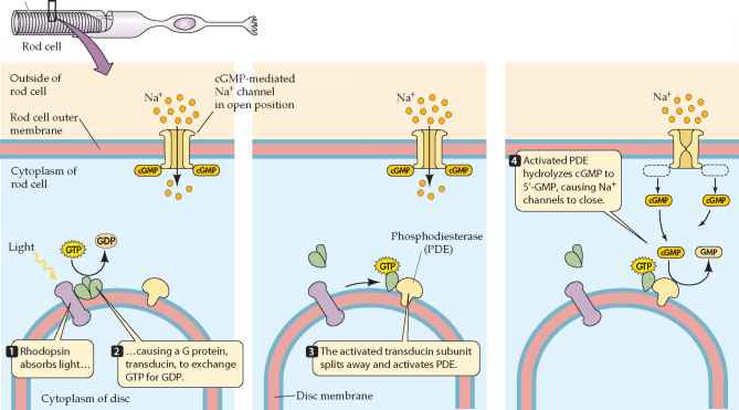
1. Photochemical activity in rods:
- Each eye contains 125 million rods which are located in neuro-retina.
- Rods contains light sensitive pigment-rhodopsin.
- Rhodopsin is a molecule formed by combination of a protein scotopsin and a light sensitive small molecule retinal (retinene).
- Retinene (retinal) is a carotenoid molecule and is derivative of vitamin A (retinol).
- Retinal exists in two isomeric form- cis and trans according to light condition.
- The extra cellular fluids surrounding rod cells contains high concentration of Na+ ion and low concentration of K+ ions while concentration of Na+ is low and K+ is high inside rod cells. The concentration is maintained by Na-K pump.
- In resting phase, K+ tends to move outside the rod cells creating slightly –ve charge inside.
- When light is falls on rod cell, it is absorbed by rhodopsin and it breaks into scotopsin and 11 cis- retinal. The process is known as bleaching.
- 11 cis-retinal absorb photon of light and change into all trans-retinal which inturn activates scotopsin into enzyme.
- This reaction produces large amount of transducin which activates another enzyme phosphodiesterase.
- Phosphodiesterase hydrolyses cGMP which causes to cease the flow of Na+ ion inside rod cell. This causes increased negative charge inside cell creating hyperpolarized state.
- Hyperpolarized rod cells transmit the neural signal to bipolar cell.
- Bipolar cell, amacrine cell and ganglion cell process the neural signal and generate action potential to transmit to brain via optic nerve.
2. Photochemical activity in cones:
- Each eye contains 7 million cone cells.
- The neural activity in cone cell is similar to that of rod cell but there are three different types of cone cells and each cone cell contains different photo-pigment and are sensitive to red, green and blue.
- Like rod, cone cell contains iodopsin as photo-pigment which is composed of 11 cis-retinal and photopsin.
- The perception of color depends upon which cone are stimulated.
- The final perceived color is combination of all three types of cone cell stimulated depending upon the level of stimulation.
- The proper mix of all three color produce the perception of white and absence of all color produce perception of black.
Processing of image in brain and perception:
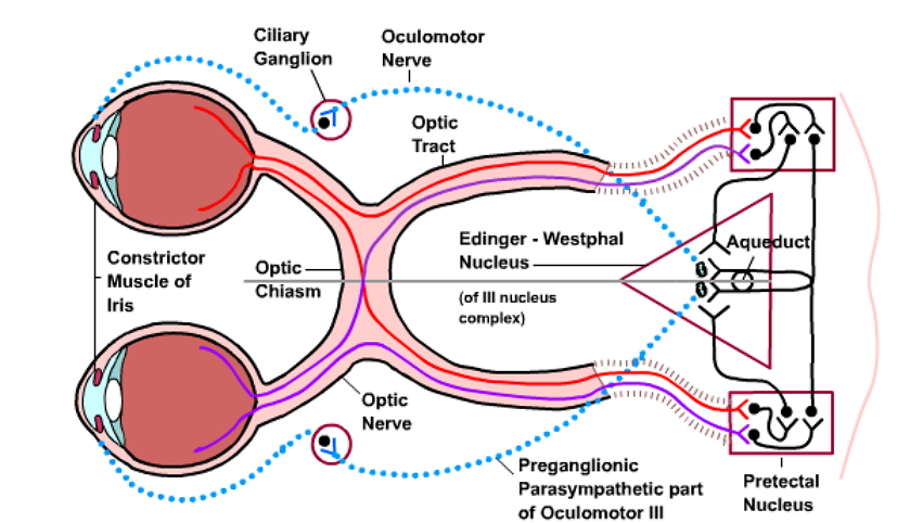
- All visual information originates in retina due to stimulation of rods and cones are conveyed to brain.
- Retina contains 5 types of cells and they are interconnected by synapse. These cells are photoreceptor cells (rod and cone), bipolar cell, ganglion cell, horizontal cell and amacrine cell.
- Photoreceptor cells, bipolar cells and ganglion cells transmit impulse directly from retina to brain.
- The nerve fiber of ganglion cells from both eyes carries impulse along two optic nerve.
- The optic nerves meets at optic chiasma where fibers from nasal half of each retina cross-over but fibers from temporal half of each retina do not cross-over.
- The optic nerve after crossing the chiasma is called as optic tract.
- Each optic tract continues posteriorly until it synapse with neuron in thalamus called lateral geniculate body which project to primary visual cortex in occipital lobe of cerebrum and image is perceived.