
Human Fertilization and Embryogenesis
Fertilization
- It is the process of Fusion of sperm and ovum to form Zygote. Fertilization usually take place in oviduct. Ovum is in secondary oocyte stage during fertilization. Secondary oocyte is surrounded by two layer-zona pellucida and zona reticulate. Sperm move toward the secondary oocyte and bind to the receptor on zona pellucida. After sperm enter the oocyte, the zona pellucida become fertilization membrane preventing other sperm to enter. It is the entry of sperm that stimulate second meiotic division of Oocyte to produce Ovum. Acrosome of sperm release proteolytic enzyme (Hyaluronidase) that digest the egg wall and then the pro-nucleuses fuse form zygote (2n).
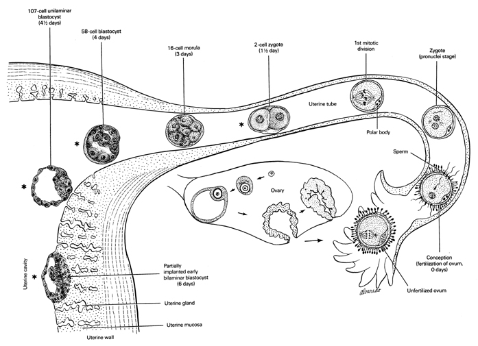
Figure: Fertilization in oviduct and Implantation of the embryo in the uterus
Embryogenesis
- Zygote undergoes repeated cell division called cleavage. Cleavage starts as the zygote moves down from oviduct to uterus 3-5 days after fertilization, zygote develop into ball like structure of cell with central cavity; blastocyst (Blastula stage).
- Outer cell of blastocyst is known as trophoblastic cell while inner cell is known as embryonic cell. Trophoblastic cell secrete HCG (human chorionic gonadotropin) hormone; similar in function as LH. It Prevent degredation of corpus luteum, therefore corpus luteum continue to secrete progesterone and oestrogen, which help continuous growth of endometrium wall causing menstruation cycle to stop.
- As blastocyst reaches to uterus, trophoblast cell invade endometrium wall and utilize nutrients for its growth and multiplication. This invasion establishes the embryo within 6-9 days in the uterus called Implantation. With successful implantation, trophoblast form chorion membrane, which later become part of placenta.
- Chorion membrane develop small villi like projection on its outer layer called Chorion villi that begins to grow in the endometrium and help in exchange of nutrition between embryo and uterus. Embryonic cell grow to become embryo. It also form other embryonic membrane covering the embryo.
- Within 20 days, embryonic membrane become clearly distinguished from embryo. The amnion is a thin membrane filled with amniotic fluids that eventually surrounds the embryo and act as shock absorbent. Later allantoin membrane develop, which develop toward the chorion and get fused to form Allanto-chorion which later form Placenta. Yolk sac has no significant function in human. Embryonic disc present between yolk sac and amnion give rise to embryo. Embryonic disc differentiate into 3 germ layer (Ectoderm, mesoderm and endoderm) known as Gastulation. Embryo shows distinct from at about 4-5 weeks. Only after 6 week, embryo can be distinguished as human embryo and now embryo term as Fetus.
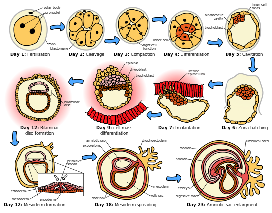
Figure: Stages of Human Embryonic development
Summary of embryonic and fetal development
- Week 1: fertilization, blastocyst formation, Implantataion
- Week2: 3 germ layer differentiate
- Week3: beginning of back bone and neural plate (first organ), embryo 2mm size long
- Week 4: heart, blood vessel, blood, gut start forming, umbilical cord develop, embryo 5mm size
- Week5: brain developing, limb buds, heart beats starts (seen on USG), embryo 8 mm long
- Week 6: eyes and ear form, embryo known as fetus
- Week 7: internal organs, face form, limbs, mouth and tongue, fetus 17mm size.
- By Week 12: fetus fully form, sex organ develop, fetus starts moving, 56mm long,
- By week 20: Hair and nails begins to grow, fingerprint develop, firm hand grip, movement of fetus can be felt, 160m long,
- By Week 24: eyelid opens, legal limit for abortion,
- By week 26: good chance of survival if prematurely born
- By week 28: respond to touch and sound, swallowing amniotic fluid, urinating
- By week 30: head lying down, 240 mm long
- Week 40: birth