
Conducting System of Heart Beat
- Heart possesses the property of autorhythmicity which means it generates it own electrical impulse and beat independent of nervous and hormonal control
- However, it is supplied with both sympathetic and para-sympathetic autonomic nerve fibres which increases or decreases the heart rate
- Heart rate is also regulated by circulating hormones eg. Adrenaline and thyroxine
- CNS exert some control over heart. However cardiac muscle of SA node initiate electric impulse for heart beat independent of CNS
- SA node is a small mass of specialized cells, that lies in the wall of right atrium near the opening of superior venacava.
- SA node is electrically unstable that generate the impulse and control its rhythm
- Since, SA node (cell of SA node) are electrically unstable, this instability leads them to discharge ( depolarize) regularly, usually 60-80 times per minute.
- Depolarizaton then transmit to adjacent muscle cells of right atrium by conduction of ions via gaps junction of intercalated discs.
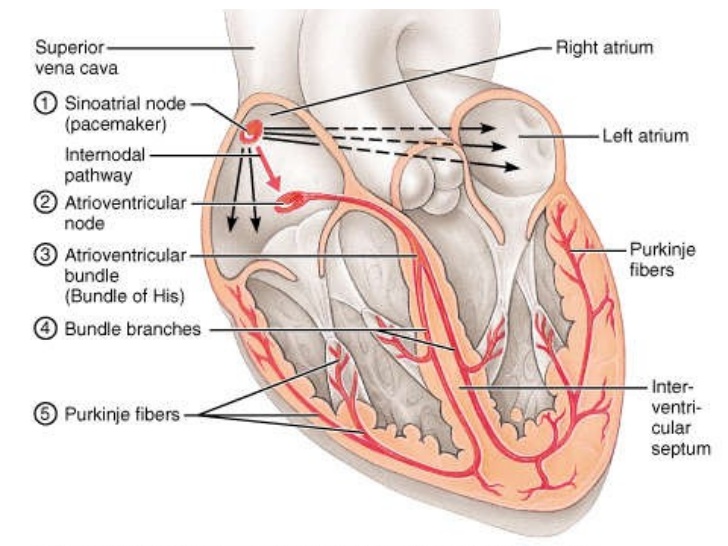 Figure: Impulse generation in SA node and transmission
 - Then atrial cells causes depolarization of neighbouring cells. Soon, the wave of electric impulse spread throughout the right and left atrium
- These electrical stimulus causes atrial contraction so that blood forced down into ventricle
- The wave of electrical impulse after leaving SA node reaches to AV node (Atrio-ventricular node)
- AV node is the small mass of neuromuscular tissue that is situated in the wall of atrial septum near Atrio-ventricular valve ( tricuspid valve)
- AV node mearly transmit the electrical signal from atrial into ventricle. There is a delay of 0.1 second for the signal to pass to ventricle.
- This delay in signal allows the atria to finish its contraction before ventricular contraction starts.
- AV node also has a secondary pacemaker function and can take over this role if there is problem with SA node for impulse generation or transmission.
- AV node passes the electrical impulse to a group of conducting fiber called Bundle of HIS (AV bundle).
- Bundle of HIS is specialized conducting fibers originates from AV node and it move downward toward the ventricle.
- Bundle of HIS divides into right and left branches as soon as it reaches inter-ventricular septum.
- Within the ventricular myocardium, bundle of HIS divides into fine fibers called purkinje’s fibers.
- AV node, AV bundle and Purkinje fibers transmit impulse throughout the ventricle causing ventricular contraction.
Mechanism of Impulse Conduction in Heart
- For the electrical conduction of heart, specialized action potential in heart is generated called Cardiac action potential.
- Cardiac action potential is divided into 5 phases.
- Resting potential
- Depolarization
- Early repolarization
- Plateau
- Repolarization
Resting potential
- In this phase, the membrane potential of cardiac muscle fiber is -90mV and these cells are stable under normal condition.
- When electrical impulses from SA node transmit, they depolarize to generate action potential.
- SA node depolarize regularly without any external influences and the depolarization transmitted to atrial cells
- Cardiac muscles fiber (cardiac myocytes) has a negative membrane potential at rest (-90mV) as like skeletal myocytes
- But in cardiac myocytes release of Ca++ ion from sarcoplasmic reticulum is induced by influx of Ca++ ion into the cell through the voltage gated calcium channel. This phenomenon is called Calcium induced calcium release.
- Increase in myoplasmic free Ca++ ion causes muscle contraction.
Depolarization
- In this phase, there is large increase in inward movement of Na+ ion through fast voltage gated Na-channel
- Increase in Na+ ion inside cell reverse the membrane potential of -90mV at rest to +30mV
- At the same time the permeability to K+ ion decreases as potassium-channel closes. So that very few K+ leaves the cell.
- This increase in Na+ ion concentration inside cell and decrease K+ ion concentration outside cell causes depolarization
Early repolarization
- In this phase, inactivation of Na-chanel occur due to influx of Ca++ ions through slow voltage gated calcium-channel.
- Influx of Ca++ causes release of Ca++ from sarcoplasmic reticulum
- Increase Ca++ ion inside cell initiate muscle contraction. And it also opens the potassium –channel, so K+ come out of the cell
Plateau
- In this phase, action potential of +30mV is maintained due to balance between inward movement of ca++ and outward movement of K+
- The Na-Ca exchanger current and Na+/K+ pumps current also play minor role during this phase.
- The balanced movement of ions (Ca++ and K+) prevents repolarization. This delay in time is called refractory period.
- The refractory period is of 0.25 seconds, which is 9 times longer than skeletal muscle.
- This extra time allow heart to refill and ensure noextra beat will occur.
Repolarization
- In this pahse, potassium- channel reopens and Calcium- channel slowly closes so that K+ move out of the cell but inward movement of Ca++ ion is blocked.
- Outward movement of K+ ion causes increase in Negative charge inside cell.
- Increase negativity inside cell returns the membrane potential to its normal -90mV. But distribution of ions are altered as there is more K+ ions outside and more Na+ ion inside cell.
- Hence, Na+ ions are pumped out of the cell and K+ ions are pumped inside cell by active transport mechanism (Na-K pump) and membrane potential is maintained at -90mv in resting phase.